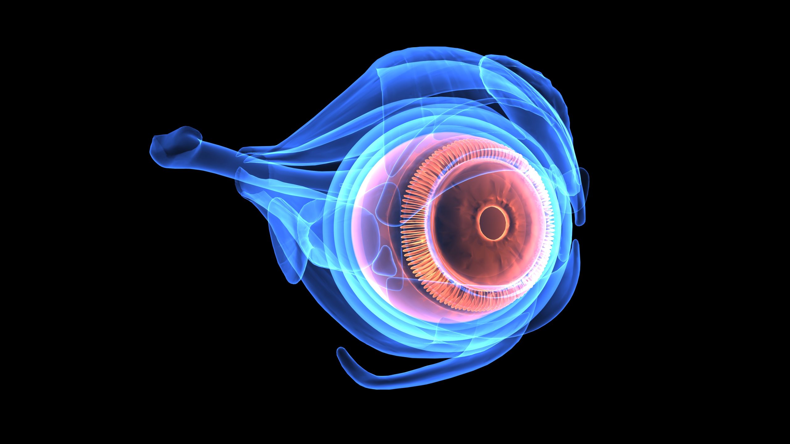
4D Imaging Technology for Real-Time Visualization of Eye Anatomy
Introduction
In the ever-evolving landscape of ophthalmology, advancements in imaging technology have been nothing short of revolutionary. One such innovation, 4D imaging technology, is poised to redefine the way we understand and treat various eye conditions. By offering real-time visualization of eye anatomy in three dimensions, along with the element of time, this cutting-edge technology holds the promise of enhancing diagnostic accuracy, surgical precision, and patient outcomes.
Understanding 4D Imaging Technology
Traditionally, ophthalmologists have relied on a combination of two-dimensional imaging techniques such as optical coherence tomography (OCT) and fundus photography to assess the structure and function of the eye. While these methods have proven invaluable, they often provide static snapshots of the eye at a single point in time, limiting our ability to capture dynamic changes and interactions within the ocular tissues.
Enter 4D imaging technology, which adds the dimension of time to the existing three-dimensional spatial information. By employing sophisticated imaging modalities and advanced computational algorithms, 4D imaging systems can generate dynamic, high-resolution images that capture the motion and kinetics of various ocular structures. This real-time visualization offers unprecedented insights into the physiological processes occurring within the eye, allowing clinicians to detect abnormalities, track disease progression, and evaluate treatment responses more effectively.
Applications in Ophthalmology
The applications of 4D imaging technology in ophthalmology are vast and diverse, spanning both diagnostic and therapeutic domains. One of its primary uses lies in the assessment of complex ocular conditions such as glaucoma, retinal diseases, and corneal disorders. By observing the dynamic behavior of tissues in real-time, clinicians can identify subtle changes indicative of disease onset or progression, enabling early intervention and tailored treatment strategies.
Moreover, 4D imaging plays a crucial role in guiding surgical interventions, particularly in procedures involving delicate ocular structures. For instance, in cataract surgery, real-time visualization of lens dynamics and intraocular fluid dynamics can aid surgeons in optimizing incision placement, phacoemulsification techniques, and intraocular lens selection, leading to improved surgical outcomes and reduced complication rates.
Advantages Over Conventional Imaging
Compared to conventional imaging modalities, 4D imaging technology offers several distinct advantages. Firstly, it provides a more comprehensive and dynamic representation of ocular anatomy and physiology, allowing for a deeper understanding of disease mechanisms and treatment responses. Secondly, by capturing real-time data, it enables clinicians to make more informed decisions in a timely manner, thereby enhancing patient care and satisfaction.
Furthermore, 4D imaging facilitates the integration of artificial intelligence (AI) algorithms for automated image analysis and diagnostic assistance. Machine learning algorithms trained on vast datasets of dynamic ocular images can help identify patterns, predict disease progression, and personalize treatment plans, ultimately optimizing clinical workflows and outcomes.
Future Directions and Challenges
While 4D imaging technology holds immense promise for the future of ophthalmology, several challenges remain to be addressed. Technical hurdles such as achieving higher spatial and temporal resolutions, reducing imaging artifacts, and enhancing image reconstruction algorithms are ongoing areas of research and development.
Additionally, the integration of 4D imaging into routine clinical practice requires overcoming logistical barriers such as cost, accessibility, and training. Collaborative efforts between researchers, clinicians, industry partners, and regulatory agencies will be essential to address these challenges and ensure widespread adoption of this transformative technology.
Conclusion
In conclusion, 4D imaging technology represents a paradigm shift in the field of ophthalmology, offering unprecedented insights into the dynamic nature of ocular anatomy and pathology. By combining spatial and temporal information in real-time, this innovative approach has the potential to revolutionize diagnostic and therapeutic strategies, ultimately improving patient outcomes and advancing the frontiers of vision health. As research continues to evolve and technologies mature, the future of ophthalmology looks brighter than ever before, illuminated by the transformative power of 4D imaging.
World Eye Care Foundation’s eyecare.live brings you the latest information from various industry sources and experts in eye health and vision care. Please consult with your eye care provider for more general information and specific eye conditions. We do not provide any medical advice, suggestions or recommendations in any health conditions.
Commonly Asked Questions
As technology continues to evolve, the future holds promise for even greater advancements in 4D imaging, paving the way for more precise diagnostics, personalized treatments, and improved outcomes for patients with eye conditions.
Absolutely, 4D imaging enables researchers to study ocular physiology, disease mechanisms, and treatment responses in unprecedented detail, advancing our understanding of vision health.
Yes, 4D imaging modalities are non-invasive and have been proven to be safe for patients of all ages, with minimal risk of adverse effects.
By visualizing retinal dynamics in real-time, 4D imaging helps clinicians monitor disease progression, assess treatment efficacy, and customize therapeutic approaches, leading to better visual outcomes for patients.
Can 4D imaging technology be integrated with artificial intelligence (AI) for diagnostic assistance?
Yes, machine learning algorithms trained on dynamic ocular images can assist in automated image analysis, pattern recognition, disease prediction, and treatment planning.
Technical challenges include achieving higher spatial and temporal resolutions, reducing imaging artifacts, and enhancing image reconstruction algorithms. Logistical challenges include cost, accessibility, and training.
Real-time visualization of lens dynamics and intraocular fluid dynamics helps surgeons optimize incision placement, phacoemulsification techniques, and intraocular lens selection, leading to improved surgical outcomes.
Yes, by capturing dynamic changes within the eye, 4D imaging can facilitate early detection and intervention for conditions such as glaucoma, retinal diseases, and corneal disorders.
4D imaging is used for diagnosing complex eye conditions, guiding surgical interventions, and monitoring disease progression and treatment responses.
4D imaging adds the dimension of time to the existing three-dimensional spatial information, enabling real-time visualization of ocular anatomy and physiology.
news via inbox
Subscribe here to get latest updates !


