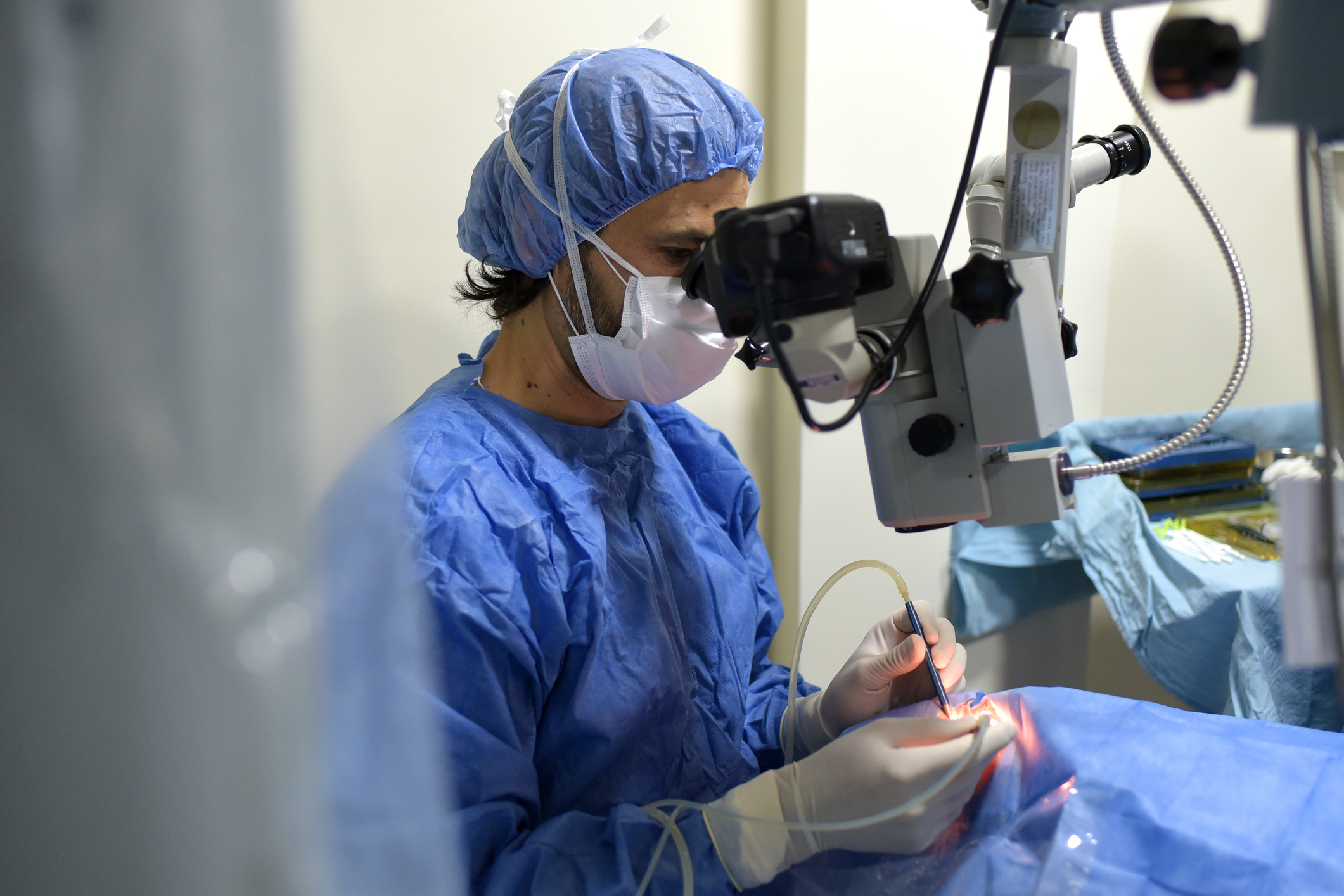
Surgical Management of Conjunctival Lesions
Introduction
Conjunctival lesions encompass a diverse range of abnormalities affecting the conjunctiva, the thin, transparent membrane covering the white part of the eye. While many conjunctival lesions are benign, some may be precancerous or malignant, necessitating surgical intervention for management. This article aims to explore the surgical approaches and considerations involved in the management of conjunctival lesions, highlighting techniques employed by ophthalmic surgeons to ensure optimal outcomes for patients.
Types of Conjunctival Lesions
Conjunctival lesions can include a variety of conditions such as:
- Pinguecula and pterygium: Non-cancerous growths often caused by exposure to ultraviolet light and environmental irritants.
- Conjunctival nevi: Benign pigmented lesions.
- Papillomas: Benign growths characterized by finger-like projections.
- Conjunctival intraepithelial neoplasia (CIN) or dysplasia: Precancerous lesions that may progress to squamous cell carcinoma.
- Squamous cell carcinoma: A malignant tumor originating from the conjunctival epithelium.
Surgical Techniques for Conjunctival Lesions
- Excisional Biopsy: This technique involves the complete removal of the conjunctival lesion along with a margin of healthy tissue. It is often used for suspicious or potentially malignant lesions to obtain a tissue sample for histopathological examination. The goal is to ensure the complete excision of abnormal cells while minimizing the risk of leaving behind residual disease. Excisional biopsy is typically performed under local anesthesia, and the excised tissue is sent to a pathology laboratory for evaluation.
- Mohs Micrographic Surgery: Mohs surgery is a specialized technique used for the precise removal of complex or recurrent lesions with indistinct margins. It involves sequential tissue excision and immediate microscopic examination of each layer to ensure complete removal while sparing healthy tissue. Mohs surgery offers the advantage of high cure rates and tissue preservation, making it particularly suitable for lesions located in cosmetically sensitive or functionally important areas, such as the eyelids or around the eye.
- Cryotherapy: Cryotherapy, or freezing, may be employed as an adjunctive treatment following surgical excision to target residual or recurrent lesions. Liquid nitrogen or a similar cryogenic agent is applied to the affected area, causing destruction of abnormal cells through rapid freezing and thawing. Cryotherapy is effective for treating superficial lesions and can help prevent recurrence by eliminating residual tumor cells. It is often used in combination with other surgical techniques to ensure comprehensive treatment.
- Amniotic Membrane Transplantation: In cases where extensive conjunctival tissue is removed, such as in the treatment of large lesions or recurrent pterygium, amniotic membrane transplantation may be utilized to promote epithelial healing and reduce scarring. Amniotic membrane, derived from the innermost layer of the placenta, possesses unique properties that facilitate tissue regeneration and modulate inflammation. It is often used as a biological scaffold to support tissue repair and promote ocular surface reconstruction following surgery.
- Autologous Conjunctival Grafting: For large defects resulting from extensive lesion removal, autologous conjunctival grafts harvested from the patient’s own conjunctiva may be utilized to reconstruct the ocular surface and maintain structural integrity. Conjunctival grafts are carefully harvested and transplanted to the site of the defect, where they provide a source of healthy tissue for epithelialization and prevent complications such as conjunctival scarring or symblepharon formation.
Considerations for Surgical Management
- Margin Assessment: Adequate margin assessment is crucial to ensure complete excision of malignant or precancerous lesions, reducing the risk of recurrence. Surgeons must carefully evaluate the surgical margins to determine the presence of residual disease and adjust the extent of excision accordingly. Margin assessment may be facilitated by intraoperative frozen section analysis or postoperative histopathological examination of the excised tissue.
- Ocular Surface Reconstruction: Preservation of ocular surface integrity is essential to maintain visual function and prevent complications such as dry eye syndrome or corneal exposure. Surgeons must employ techniques to promote epithelial healing and prevent adhesions or contractures that may compromise ocular mobility. Strategies for ocular surface reconstruction may include the use of amniotic membrane, conjunctival grafts, or tissue adhesives to facilitate wound closure and promote tissue regeneration.
- Long-term Follow-up: Regular postoperative monitoring is essential to detect recurrence or complications early and ensure optimal outcomes for patients undergoing surgical management of conjunctival lesions. Patients should be scheduled for follow-up appointments at regular intervals to assess healing, monitor for signs of recurrence, and address any postoperative complications promptly. Long-term surveillance is particularly important for patients with a history of malignancy or high-risk lesions to detect recurrence or progression early and intervene as necessary. Close collaboration between ophthalmologists, pathologists, and other members of the healthcare team is essential to ensure comprehensive care and long-term success in the management of conjunctival lesions.
Conclusion
Surgical management plays a pivotal role in the treatment of conjunctival lesions, ranging from benign growths to malignant tumors. By employing a combination of excisional techniques, adjuvant therapies, and meticulous surgical planning, ophthalmic surgeons can effectively manage conjunctival lesions while preserving ocular function and optimizing patient outcomes. Close collaboration between ophthalmologists, pathologists, and other members of the healthcare team is essential to ensure comprehensive care and long-term success in the management of conjunctival lesions.
World Eye Care Foundation’s eyecare.live brings you the latest information from various industry sources and experts in eye health and vision care. Please consult with your eye care provider for more general information and specific eye conditions. We do not provide any medical advice, suggestions or recommendations in any health conditions.
Commonly Asked Questions
Individuals with a history of conjunctival lesions should undergo regular eye examinations as recommended by their ophthalmologist. This frequency may vary depending on the type and severity of the lesions and individual risk factors.
Yes, conjunctival lesions such as pinguecula and pterygium can cause symptoms like redness, irritation, and foreign body sensation, especially if they grow large enough to encroach on the cornea.
Depending on the type and severity of the lesion, non-surgical treatments such as topical medications or cryotherapy may be considered as alternatives to surgery.
While some conjunctival lesions may be preventable by avoiding excessive UV exposure and protecting the eyes from environmental irritants, others may develop due to factors beyond one’s control.
Risk factors for conjunctival lesions include prolonged exposure to ultraviolet light, environmental irritants, history of eye trauma, and certain genetic predispositions.
Yes, certain types of conjunctival lesions, particularly precancerous or malignant ones, may recur after surgical removal. Regular follow-up visits with an ophthalmologist are essential for monitoring and early detection of recurrence.
Recovery time can vary depending on the type and extent of surgery performed. Most patients can expect improvement within a few weeks, but complete healing may take several months.
No, many conjunctival lesions are benign, such as pinguecula and pterygium. However, some lesions, such as squamous cell carcinoma, can be malignant and require prompt treatment.
Surgical removal is a common treatment for conjunctival lesions, but other options such as cryotherapy or topical medications may be considered depending on the type and severity of the lesion.
In some cases, conjunctival lesions may cause vision disturbances if they affect the cornea or obstruct the visual axis. However, not all conjunctival lesions directly impact vision.
news via inbox
Subscribe here to get latest updates !


