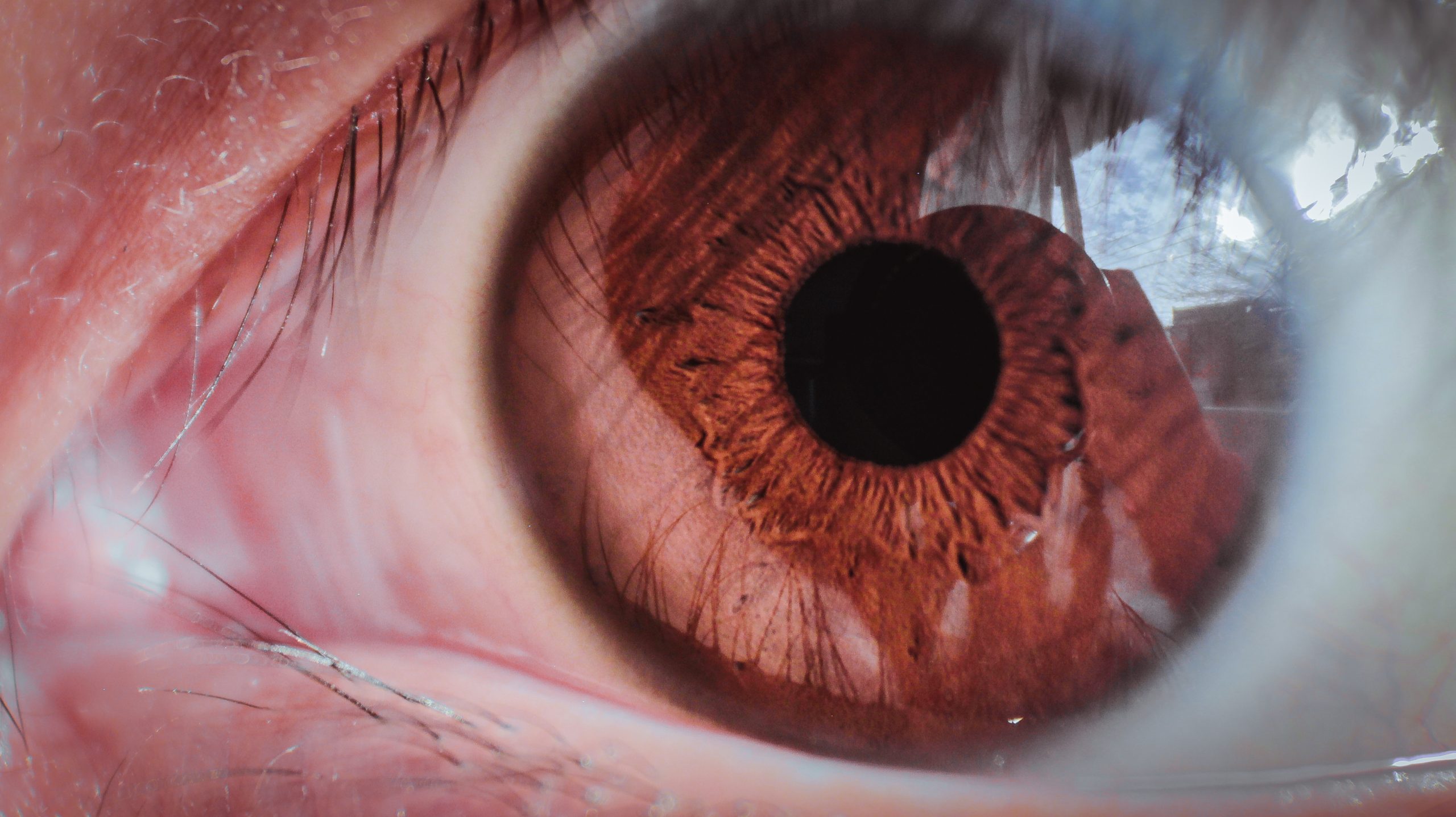
A Comprehensive Guide to Primary Congenital Glaucoma
Primary congenital glaucoma, often referred to as infantile glaucoma or congenital glaucoma, is a rare but serious eye condition that manifests in infancy or early childhood. Unlike adult-onset glaucoma, which often results from aging and other systemic factors, primary congenital glaucoma is caused by developmental abnormalities in the eye’s drainage structures. It represents a subset of glaucoma disorders that affect the eyes from birth or shortly thereafter. Despite its rarity, primary congenital glaucoma demands attention due to its potential to cause irreversible vision loss if left untreated. In this article, we delve into the intricacies of this condition, exploring its causes, symptoms, diagnosis, and treatment options.
Understanding Primary Congenital Glaucoma
Primary congenital glaucoma stems from abnormalities in the eye’s drainage system, specifically the trabecular meshwork—a network of tissues responsible for maintaining the balance of intraocular fluid. In affected individuals, this drainage system fails to function properly, leading to impaired fluid outflow and subsequent elevation of intraocular pressure (IOP). Elevated IOP places undue stress on the delicate optic nerve, which transmits visual information from the eye to the brain. Over time, this pressure can cause irreversible damage to the optic nerve fibers, resulting in vision loss if left untreated. While primary congenital glaucoma is relatively rare, affecting approximately 1 in every 10,000 births, its potential impact on vision underscores the importance of early detection and intervention.
Signs and Symptoms
Recognizing the signs of primary congenital glaucoma is crucial for timely diagnosis and management. Although infants and young children may not be able to articulate their symptoms, parents and caregivers can be vigilant for the following indicators:
- Excessive tearing: Infants with primary congenital glaucoma often exhibit persistent tearing, which may be mistaken for normal tearing associated with teething or other benign causes. However, if tearing persists beyond the first few months of life, it could signal a blocked or malfunctioning tear drainage system—a hallmark feature of congenital glaucoma.
- Photophobia: Sensitivity to light is another common symptom of primary congenital glaucoma. Affected infants may squint or shield their eyes in response to bright light, as excessive light exposure can exacerbate discomfort and visual disturbances.
- Enlarged eyes: Buphthalmos, or enlargement of one or both eyes, is a characteristic feature of primary congenital glaucoma. This enlargement results from the accumulation of fluid within the eye, leading to stretching and expansion of the ocular tissues. In severe cases, the affected eye may appear significantly larger than the unaffected eye, a condition known as megalophthalmos.
- Corneal cloudiness: Corneal edema, or cloudiness of the cornea, can occur secondary to elevated intraocular pressure in primary congenital glaucoma. The cornea, which is normally transparent, may become hazy or opaque due to fluid buildup, impairing visual acuity and clarity.
In addition to these primary symptoms, infants with congenital glaucoma may also exhibit irritability, poor feeding, and decreased responsiveness, reflecting their discomfort and visual impairment. It’s important for parents to consult a pediatric ophthalmologist if they observe any of these signs in their child, as early intervention can help preserve vision and prevent long-term complications.
Diagnosis and Evaluation
Diagnosing primary congenital glaucoma requires a thorough clinical evaluation by a skilled ophthalmologist, preferably one with expertise in pediatric eye care. The diagnostic process typically includes the following steps:
- Comprehensive eye examination: The ophthalmologist will conduct a detailed assessment of the child’s ocular structures, including the cornea, iris, lens, and optic nerve. Special attention will be given to the appearance of the cornea, which may exhibit signs of edema, and the size of the eye, which may be enlarged in cases of buphthalmos.
- Measurement of intraocular pressure: Using a handheld tonometer or a specialized device, the ophthalmologist will measure the intraocular pressure to assess for elevated levels indicative of glaucoma. In infants, this measurement may be performed while the child is asleep to minimize discomfort and ensure accuracy.
- Evaluation of the drainage angle: The drainage angle, where the aqueous humor exits the eye, is examined to determine if there are any structural abnormalities or blockages impeding fluid outflow. This assessment helps guide treatment decisions and prognostic considerations.
- Imaging studies: In some cases, imaging modalities such as ultrasound biomicroscopy or optical coherence tomography (OCT) may be utilized to visualize the internal structures of the eye in greater detail. These non-invasive techniques provide valuable insights into the anatomy and pathology associated with primary congenital glaucoma.
By combining these diagnostic tools and techniques, ophthalmologists can establish an accurate diagnosis of primary congenital glaucoma and develop a tailored treatment plan to address the child’s needs.
Treatment Approaches
The management of primary congenital glaucoma aims to reduce intraocular pressure, preserve optic nerve function, and optimize visual outcomes. Treatment options may include:
- Medication: Topical or oral medications may be prescribed to lower intraocular pressure by either increasing the outflow of aqueous humor or decreasing its production within the eye. Common medications used in the treatment of congenital glaucoma include prostaglandin analogs, beta-blockers, and carbonic anhydrase inhibitors. These medications are often administered multiple times per day and require careful monitoring of efficacy and side effects.
- Surgical intervention: When medical therapy alone is insufficient to control intraocular pressure, surgical intervention may be necessary to restore normal drainage function. Several surgical techniques may be employed, including:
- Trabeculotomy: A microsurgical procedure in which the trabecular meshwork is incised or removed to improve aqueous outflow.
- Trabeculectomy: The creation of a surgical fistula or bypass channel to facilitate drainage of aqueous humor from the eye, often combined with the placement of an absorbable or permanent drainage implant.
- Goniotomy: A minimally invasive procedure in which a specialized lens is used to visualize and incise the trabecular meshwork, enhancing drainage efficiency.
- Tube shunt implantation: Placement of a small tube or shunt device within the eye to divert aqueous humor to an external reservoir, reducing intraocular pressure.
The choice of surgical technique depends on various factors, including the severity of glaucoma, the age of the child, and the presence of concurrent ocular or systemic conditions. Surgical interventions for congenital glaucoma are typically performed under general anesthesia to ensure the comfort and safety of the child.
Long-Term Management and Prognosis
Successful management of primary congenital glaucoma requires ongoing monitoring and follow-up care to assess treatment efficacy, detect disease progression, and address any complications that may arise. Children diagnosed with congenital glaucoma may require lifelong management to maintain optimal intraocular pressure and preserve visual function.
Regular eye examinations, including measurement of intraocular pressure and evaluation of optic nerve health, are essential components of long-term management. Additionally, parents and caregivers should be educated about the signs and symptoms of glaucoma recurrence or complications, such as elevated IOP, corneal scarring, or refractive errors, which may necessitate adjustments to treatment or additional interventions.
Despite the challenges posed by primary congenital glaucoma, early diagnosis and intervention can significantly improve the prognosis and quality of life for affected children. With advances in medical and surgical techniques, coupled with comprehensive multidisciplinary care, many children with congenital glaucoma can achieve favorable visual outcomes and lead fulfilling lives.
Conclusion
Primary congenital glaucoma represents a complex and potentially sight-threatening condition that requires prompt recognition and intervention. By understanding the signs, symptoms, and treatment options associated with congenital glaucoma, parents, caregivers, and healthcare providers can work together to ensure early diagnosis, effective management, and optimal visual outcomes for affected children. Through continued research, education, and advocacy efforts, we can strive to enhance awareness, improve access to care, and ultimately, mitigate the impact of congenital glaucoma on individuals and families worldwide.
World Eye Care Foundation’s eyecare.live brings you the latest information from various industry sources and experts in eye health and vision care. Please consult with your eye care provider for more general information and specific eye conditions. We do not provide any medical advice, suggestions or recommendations in any health conditions.
Commonly Asked Questions
Risk factors for primary congenital glaucoma include a family history of the condition, genetic mutations, and certain developmental abnormalities of the eye’s drainage structures.
While prenatal screening for congenital glaucoma is not routine, certain imaging techniques such as ultrasound may detect structural abnormalities suggestive of the condition in utero.
Yes, primary congenital glaucoma can have a hereditary component, with mutations in genes such as CYP1B1 and FOXC1 implicated in its development. However, not all cases are inherited, and spontaneous mutations can also occur.
While lifestyle modifications alone are not sufficient to treat primary congenital glaucoma, maintaining a healthy lifestyle, including regular exercise and a balanced diet, may support overall ocular health and complement medical or surgical interventions.
Yes, primary congenital glaucoma can affect one or both eyes, though it may be asymmetrical in some cases. Bilateral involvement is relatively common, highlighting the importance of comprehensive evaluation and treatment for both eyes.
Primary congenital glaucoma is typically diagnosed within the first year of life, with symptoms often manifesting in the first few months after birth. Early detection and intervention are critical for preserving vision and preventing complications.
While primary congenital glaucoma is not curable in the traditional sense, timely diagnosis and appropriate management can help control intraocular pressure, preserve optic nerve function, and optimize visual outcomes. With effective treatment, many individuals with congenital glaucoma can lead healthy, fulfilling lives.
Untreated primary congenital glaucoma can lead to irreversible vision loss, corneal scarring, amblyopia (lazy eye), and other ocular complications. Early intervention is essential to minimize the risk of long-term sequelae.
While treatment for primary congenital glaucoma can be effective in controlling intraocular pressure, the condition may recur or progress over time, necessitating ongoing monitoring and adjustments to management strategies as needed.
Families affected by primary congenital glaucoma can benefit from support groups, educational materials, and advocacy organizations dedicated to raising awareness, providing information, and connecting individuals with resources and services tailored to their needs.
news via inbox
Subscribe here to get latest updates !


