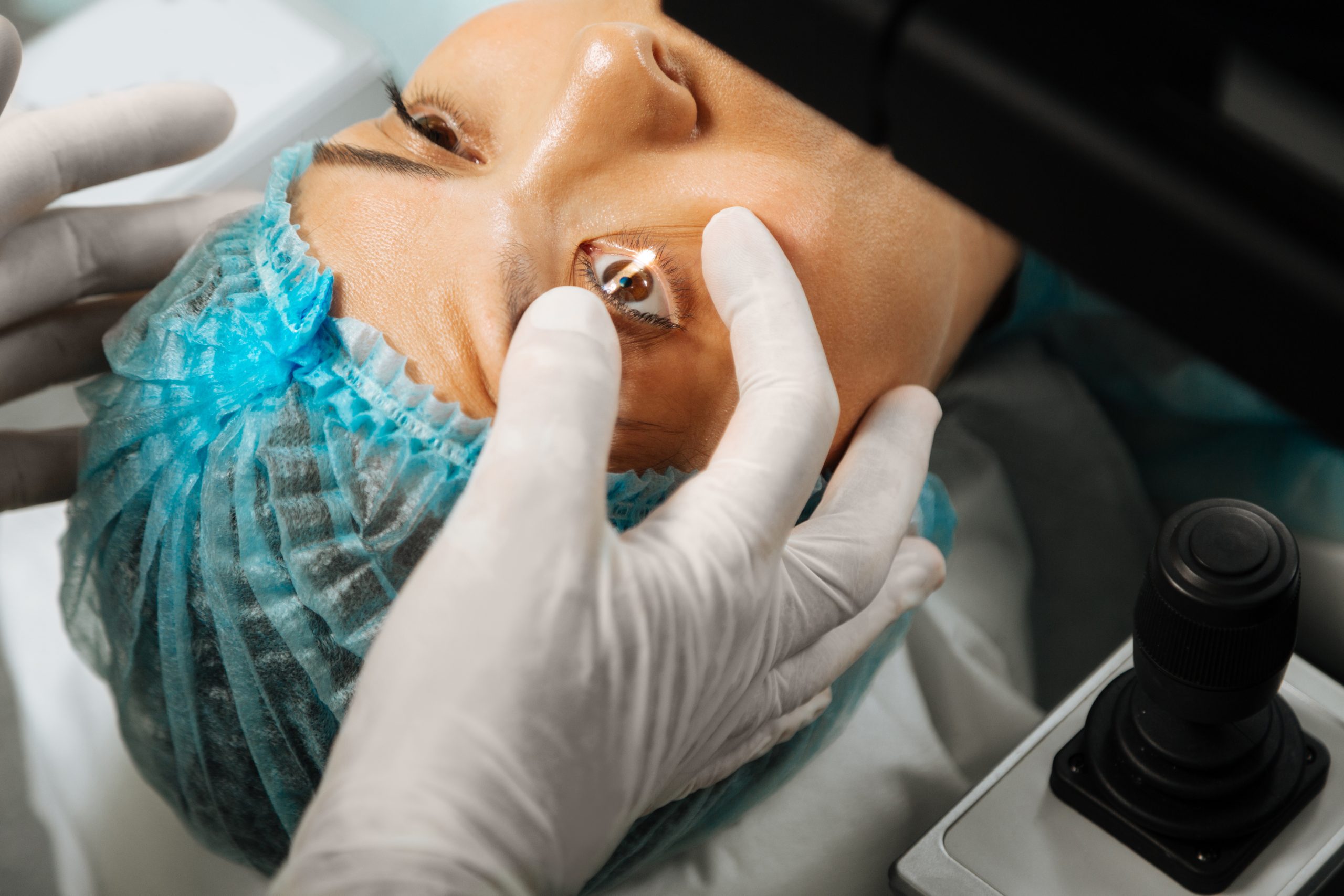
Understanding Deep Anterior Lamellar Keratoplasty (DALK)
Introduction
Deep Anterior Lamellar Keratoplasty (DALK) stands as a revolutionary surgical technique in the realm of ophthalmology, offering renewed hope for individuals suffering from corneal diseases or injuries. This intricate procedure targets the cornea, the transparent front part of the eye responsible for refracting light onto the retina, thus enabling vision. DALK aims to restore vision by selectively replacing damaged or diseased corneal tissue while preserving the endothelium, the delicate innermost layer crucial for maintaining corneal clarity. Let’s delve into the depths of DALK, exploring its techniques, benefits, risks, and post-operative care.
What is DALK?
DALK involves the partial removal and replacement of the anterior layers of the cornea, leaving the endothelium intact. This crucial distinction from traditional penetrating keratoplasty (PKP), where all layers of the cornea are replaced, mitigates the risk of endothelial rejection and associated complications. DALK is primarily indicated for conditions affecting the corneal stroma, such as keratoconus, corneal dystrophies, and scars resulting from trauma or infections.
The DALK Procedure
DALK surgery comprises several key steps, each meticulously executed to ensure optimal outcomes:
- Corneal Dissection: The initial step in DALK involves creating an entry point into the cornea, typically at the peripheral region. This incision provides access for subsequent dissection and graft placement.
- Manual or Automated Dissection: Depending on the surgeon’s preference and patient’s condition, the stromal layers are separated either manually using specialized instruments or utilizing advanced femtosecond laser technology. Manual dissection involves the use of instruments like a crescent blade or a microkeratome to create a partial-thickness trephination, followed by careful separation of stromal layers using dissecting instruments such as a spatula or a big bubble technique. Femtosecond laser-assisted DALK offers precise, bladeless dissection, enhancing safety and reproducibility.
- Removal of Diseased Tissue: Once the stromal layers are adequately dissected, the targeted layers of the cornea, including the diseased or scarred tissue, are delicately excised. This step requires meticulous attention to avoid damage to the underlying Descemet’s membrane and endothelium.
- Graft Preparation and Placement: The donor corneal tissue, obtained from a cadaveric source or through eye banking, is meticulously prepared to match the recipient’s size and shape. The endothelial layer is typically removed to minimize the risk of rejection. The graft is then carefully positioned and secured onto the host bed using sutures or tissue adhesive.
- Suturing or Adhesive Application: Sutures may be utilized to secure the graft in place, ensuring proper apposition and stability during the initial healing phase. Alternatively, tissue adhesive, such as fibrin glue, may be employed to facilitate faster healing and reduce the risk of induced astigmatism. The choice between sutures and adhesive depends on the surgeon’s preference, graft-host interface quality, and patient factors.
Benefits of DALK
DALK offers several advantages over traditional full-thickness corneal transplantation, including:
- Reduced Risk of Rejection: By preserving the endothelium, DALK significantly lowers the risk of immune-mediated rejection, leading to improved graft survival rates. The absence of endothelial rejection also reduces the need for long-term immunosuppressive therapy, minimizing systemic side effects.
- Enhanced Visual Outcomes: With precise removal of diseased tissue and meticulous graft placement, DALK often results in superior visual acuity and reduced post-operative astigmatism. Studies have demonstrated comparable or even better visual outcomes with DALK compared to PKP, particularly in cases of keratoconus and stromal dystrophies.
- Stable Refractive Results: The selective replacement of corneal layers minimizes changes in corneal curvature, providing more predictable and stable refractive outcomes. This stability is particularly advantageous for patients with pre-existing refractive errors or those seeking corneal transplantation for visual rehabilitation.
- Faster Recovery: Compared to PKP, DALK typically entails a shorter recovery period, allowing patients to resume normal activities sooner. The preservation of corneal nerves and reduced disruption of ocular surface integrity contribute to quicker visual rehabilitation and less post-operative discomfort.
Risks and Considerations
While DALK offers promising outcomes, it is not without potential risks and considerations:
- Incomplete Dissection: Inadequate separation of stromal layers can complicate graft placement and increase the risk of intraoperative complications, such as Descemet’s membrane perforation or inadvertent endothelial damage. Surgeon experience and familiarity with various dissection techniques are crucial for minimizing this risk.
- Interface Opacities: The interface between the donor and recipient corneal tissue may sometimes develop opacities, affecting visual clarity. These interface opacities can arise due to irregular graft-host apposition, interface debris, or interface inflammation. Close post-operative monitoring and timely intervention, such as selective epithelial removal or interface irrigation, may be necessary to address interface opacities and optimize visual outcomes.
- Steroid-Induced Glaucoma: Prolonged use of corticosteroid medications, essential for preventing graft rejection, may predispose patients to elevated intraocular pressure and glaucoma. Regular monitoring of intraocular pressure and judicious steroid tapering are essential for early detection and management of steroid-induced ocular hypertension.
- Graft Failure: Despite meticulous surgical technique, graft failure remains a possibility, necessitating close monitoring and potential re-intervention. Graft failure may result from various factors, including immune-mediated rejection, graft infection, or endothelial decompensation. Prompt recognition of graft failure signs, such as persistent epithelial defects, corneal edema, or graft vascularization, allows for timely intervention, which may involve repeat DALK, conversion to PKP, or alternative treatment modalities such as Descemet membrane endothelial keratoplasty (DMEK).
Post-Operative Care
Following DALK surgery, patients require diligent post-operative care to promote optimal healing and minimize complications. This typically includes:
- Topical Medications: Patients are prescribed a regimen of antibiotic and corticosteroid eye drops to prevent infection and inflammation. The frequency and duration of topical medications may vary depending on the surgeon’s protocol and the patient’s individual response.
- Regular Follow-Up Visits: Scheduled follow-up appointments allow the surgeon to monitor graft healing, assess visual acuity, and adjust medications as needed. Early post-operative visits are crucial for evaluating graft clarity, detecting signs of rejection or infection, and addressing any concerns or symptoms reported by the patient.
- Eye Protection: Patients are advised to avoid rubbing or exerting pressure on the eyes and to use protective eyewear, especially during activities that pose a risk of trauma. Compliance with post-operative restrictions, such as avoiding strenuous activities, swimming, or exposure to dusty or windy environments, minimizes the risk of post-operative complications and promotes optimal graft integration.
- Patience and Compliance: Full visual recovery may take several months, and strict adherence to post-operative instructions is paramount for optimal outcomes. Patients should be counseled on the expected timeline for visual rehabilitation, which may include temporary fluctuations in vision, gradual improvement in visual acuity, and stabilization of refractive error. Encouraging patients to adhere to their prescribed medication regimen, attend scheduled follow-up visits, and report any unusual symptoms or changes in vision facilitates early detection and management of post-operative complications, ensuring the best possible visual outcomes.
Conclusion
Deep Anterior Lamellar Keratoplasty (DALK) represents a significant advancement in corneal surgery, offering a tailored approach to sight restoration with minimized risk of rejection and improved visual outcomes. While the procedure requires precision and expertise, its potential to transform the lives of individuals affected by corneal diseases or injuries cannot be overstated. By understanding the techniques, benefits, risks, and post-operative care associated with DALK, patients and healthcare providers alike can make informed decisions and pave the way for clearer vision and enhanced quality of life. Ongoing research and technological innovations continue to refine DALK techniques and expand its applicability, further advancing the field of corneal transplantation and optimizing outcomes for patients worldwide.
World Eye Care Foundation’s eyecare.live brings you the latest information from various industry sources and experts in eye health and vision care. Please consult with your eye care provider for more general information and specific eye conditions. We do not provide any medical advice, suggestions or recommendations in any health conditions.
Commonly Asked Questions
While recovery times vary, most patients can resume light activities within a few days to weeks after DALK surgery. Strenuous activities, contact sports, and swimming should be avoided for several weeks to minimize the risk of complications and promote optimal healing.
If DALK surgery is not suitable or successful, alternative treatments may include traditional penetrating keratoplasty (PKP), Descemet membrane endothelial keratoplasty (DMEK), or innovative techniques such as corneal collagen cross-linking (CXL) for keratoconus management. The optimal treatment approach depends on the specific diagnosis, corneal characteristics, and patient preferences.
In cases of graft failure or suboptimal outcomes, repeat DALK surgery may be considered to restore vision and improve corneal health. The decision to repeat DALK depends on various factors, including the cause of graft failure, ocular surface integrity, and the patient’s overall health and visual goals.
In some cases, DALK may be feasible for patients with a history of previous eye surgeries, depending on the specific circumstances and underlying corneal pathology. However, prior surgeries may affect the complexity and outcomes of DALK, and thorough pre-operative evaluation is necessary to assess candidacy.
Suture removal timing varies depending on the patient’s healing response and graft stability. In general, sutures may be gradually removed in a staged fashion over several months to years post-operatively, guided by clinical assessment of graft integration, astigmatism, and corneal clarity.
Complications may include incomplete dissection, interface opacities, steroid-induced glaucoma, and graft failure. However, with experienced surgeons and careful patient selection, the risk of complications is minimized.
Performing DALK on both eyes simultaneously may increase the risk of post-operative complications and affect recovery. Surgeons typically recommend staged procedures to minimize potential risks and optimize outcomes.
DALK offers reduced risk of endothelial rejection compared to penetrating keratoplasty (PKP) since it preserves the recipient’s endothelium. This reduces the need for long-term immunosuppressive therapy and lowers the risk of rejection-related complications.
Recovery time varies, but patients typically experience improved vision within a few weeks to months. Full visual recovery may take several months, and adherence to post-operative care instructions is essential.
DALK is primarily indicated for corneal conditions affecting the stroma, including keratoconus, corneal dystrophies, and scars resulting from trauma or infections. It selectively replaces damaged tissue while preserving the endothelium.
news via inbox
Subscribe here to get latest updates !


