
Comprehensive Guide to Color Doppler Ultrasonography
Introduction
Color Doppler ultrasonography is a valuable diagnostic tool used in various medical fields, including ophthalmology, to assess blood flow dynamics and vascular abnormalities. This non-invasive imaging technique combines conventional ultrasound with Doppler technology to visualize and quantify blood flow in real-time. Here’s a detailed exploration of Color Doppler ultrasonography, its principles, applications, benefits, and clinical relevance:
Principles of Color Doppler Ultrasonography
Color Doppler ultrasonography combines traditional ultrasound imaging with Doppler technology to visualize and quantify blood flow dynamics. The key principles include:
- Doppler Effect: This fundamental principle involves the change in frequency of sound waves reflected from moving objects (blood cells) compared to stationary objects (tissue). By detecting these frequency shifts, Color Doppler ultrasound can calculate the velocity and direction of blood flow within vessels.
- Color Coding: In Color Doppler imaging, blood flow is represented as color-coded signals overlaid on grayscale ultrasound images. Typically, red indicates blood flow towards the transducer (positive Doppler shift), while blue indicates flow away from the transducer (negative Doppler shift). This color mapping allows for intuitive visualization of blood flow direction and speed.
- Pulsed and Continuous Wave Doppler: These are two techniques used in Color Doppler ultrasonography:
- Pulsed Doppler: Sends and receives ultrasound waves in short bursts, allowing for detailed measurement of blood flow velocities at specific depths within vessels. It’s useful for assessing flow characteristics in smaller vessels and around anatomical structures like the optic nerve.
- Continuous Wave Doppler: Uses continuous transmission and reception of ultrasound waves, enabling broader assessment of blood flow velocities across larger vessels. It’s particularly useful for measuring high velocities, such as in the central retinal artery and vein.
Applications in Ophthalmology
Color Doppler ultrasonography in ophthalmology is crucial for evaluating various conditions affecting ocular blood flow and vascular dynamics:
- Glaucoma: Studies have shown that impaired ocular blood flow may contribute to the pathogenesis of glaucoma. Color Doppler ultrasound helps measure blood flow velocities in retrobulbar vessels, including the ophthalmic artery and central retinal artery. Changes in these velocities can indicate vascular dysregulation associated with glaucomatous optic neuropathy.
- Optic Nerve Disorders: Assessing blood flow to the optic nerve head is essential for diagnosing and monitoring conditions such as optic neuritis, ischemic optic neuropathy, and papilledema. Color Doppler ultrasound provides insights into changes in blood flow velocities, helping differentiate between ischemic and non-ischemic optic nerve disorders.
- Orbital Tumors: Color Doppler imaging aids in characterizing orbital masses by distinguishing vascular tumors (e.g., hemangiomas) from non-vascular lesions (e.g., dermoid cysts). Vascular tumors typically exhibit increased vascularity and turbulent blood flow patterns, which are visualized as high-velocity signals on Doppler ultrasound.
Clinical Benefits
Color Doppler ultrasonography offers several advantages in clinical practice:
- Non-invasive and Safe: Unlike invasive angiography, Color Doppler ultrasound does not involve ionizing radiation or contrast agents, making it safe for use in patients, including pregnant women and children. It can be repeated as needed without significant risk.
- Real-time Imaging: Provides immediate visualization of blood flow dynamics in real-time, enabling clinicians to assess changes during maneuvers like ocular compression or when evaluating dynamic conditions such as arteriovenous fistulas.
- Quantitative Analysis: In addition to visualizing blood flow direction and velocity through color coding, Color Doppler ultrasound allows for quantitative measurements such as:
- Peak Systolic Velocity (PSV): Maximum velocity of blood flow during systole.
- End-Diastolic Velocity (EDV): Minimum velocity of blood flow during diastole.
- Resistive Index (RI): Calculated as (PSV – EDV) / PSV, RI provides insights into vascular resistance and is used to assess the hemodynamic significance of vascular lesions.
Procedure and Interpretation
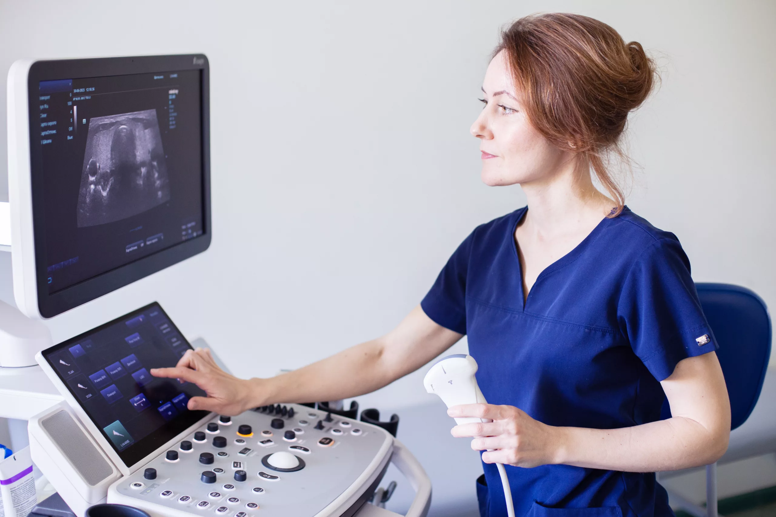
- Patient Preparation: Before the procedure, patients are positioned comfortably, usually lying down. A water-based gel is applied to the eyelid or orbital area to facilitate sound wave transmission and ensure optimal image quality.
- Image Acquisition: The ultrasound transducer is gently placed on the closed eyelid or orbital rim, using minimal pressure to avoid discomfort. The transducer emits ultrasound waves and receives reflected signals, which are processed to create real-time images of ocular structures and blood vessels.
- Interpretation: Doppler ultrasound images display color-coded blood flow patterns overlaid on grayscale anatomical images. Clinicians interpret these patterns to assess:
- Flow Direction: Red (towards the transducer) or blue (away from the transducer).
- Flow Velocity: Visualized as varying intensities of color, with higher velocities represented by brighter colors.
- Vascular Anatomy: Identification of major vessels, detection of abnormalities such as stenosis or aneurysms, and assessment of collateral circulation.
Limitations and Considerations
Despite its advantages, Color Doppler ultrasonography has limitations and considerations:
- Operator Dependence: The quality of images and accuracy of measurements depend on the operator’s experience and skill in ultrasound technique.
- Anatomical Challenges: Visualization may be limited by bone structures (e.g., orbital walls) and air interfaces (e.g., within the sinuses), particularly when assessing deeper orbital structures or posterior ocular segments.
- Artifact Interference: Patient factors such as eyelid position, patient movement, or inadequate gel application can lead to imaging artifacts, affecting image quality and diagnostic accuracy.
Future Directions and Research
Ongoing research aims to enhance the utility and accuracy of Color Doppler ultrasonography in ophthalmology:
- Technological Advancements: Continued improvements in ultrasound technology focus on enhancing spatial resolution, reducing artifact interference, and expanding capabilities for quantitative flow analysis.
- Clinical Applications: Exploring new applications, such as assessing choroidal blood flow in conditions like age-related macular degeneration (AMD) and diabetic retinopathy, to broaden the scope of diagnostic and monitoring capabilities.
- Research Initiatives: Investigating the role of Doppler parameters (e.g., pulsatility index, flow volume) as biomarkers for ocular disease progression and treatment response, facilitating personalized medicine approaches in ophthalmology.
Conclusion
Color Doppler ultrasonography is a valuable imaging modality in ophthalmology, providing essential insights into ocular blood flow dynamics and vascular pathologies. Its non-invasive nature, real-time imaging capabilities, and quantitative analysis tools make it indispensable for diagnosing and managing a wide range of ocular conditions, from glaucoma to orbital tumors. As technology advances and research progresses, Color Doppler ultrasonography continues to evolve, promising further improvements in diagnostic accuracy and patient care outcomes.
World Eye Care Foundation’s eyecare.live brings you the latest information from various industry sources and experts in eye health and vision care. Please consult with your eye care provider for more general information and specific eye conditions. We do not provide any medical advice, suggestions or recommendations in any health conditions.
Commonly Asked Questions
Color Doppler ultrasonography is commonly used to evaluate ocular blood flow in conditions such as glaucoma, optic nerve disorders (e.g., ischemic optic neuropathy), and orbital tumors. It helps assess vascular contributions to these diseases and monitor changes in blood flow dynamics over time.
No, Color Doppler ultrasonography is non-invasive and painless. It uses ultrasound waves to visualize blood flow in the eyes and orbits without the need for injections or incisions. It’s considered safe for patients of all ages, including pregnant women.
Color Doppler ultrasonography can measure blood flow velocities in retrobulbar vessels, including the ophthalmic artery and central retinal artery. Changes in these velocities may indicate vascular dysregulation associated with glaucomatous optic neuropathy, aiding in the diagnosis and management of glaucoma.
Vascular tumors, such as hemangiomas, typically exhibit increased vascularity and turbulent blood flow patterns, which appear as high-velocity signals on Color Doppler ultrasound. Non-vascular lesions, like dermoid cysts, lack such vascular flow patterns, aiding in their differentiation.
Limitations include operator dependence on technique and interpretation, challenges in visualizing deeper orbital structures due to bone and air interfaces, and potential for imaging artifacts from patient factors like eyelid position or movement during the procedure.
Can Color Doppler ultrasonography detect changes in blood flow associated with diabetic retinopathy?
Yes, Color Doppler ultrasonography can assess choroidal and retinal blood flow, providing insights into microvascular changes seen in diabetic retinopathy. It helps evaluate perfusion abnormalities and monitor response to treatment interventions.
Doppler parameters include Peak Systolic Velocity (PSV), End-Diastolic Velocity (EDV), and Resistive Index (RI). PSV and EDV measure maximum and minimum blood flow velocities, respectively, while RI indicates vascular resistance. These parameters help quantify blood flow characteristics and assess vascular health.
Yes, Color Doppler ultrasonography is safe and effective for use in pediatric patients. It provides valuable information about ocular blood flow dynamics and helps diagnose conditions such as congenital vascular anomalies or pediatric glaucoma.
Researchers are exploring new applications, such as assessing choroidal blood flow in age-related macular degeneration (AMD), evaluating optic nerve perfusion in neurodegenerative diseases, and studying vascular changes in inflammatory eye conditions. These advancements aim to expand diagnostic capabilities and refine treatment strategies.
Color Doppler ultrasonography offers real-time visualization of blood flow without the need for contrast agents used in FA or the magnetic fields used in MRA. It provides complementary information about blood flow dynamics and vascular anatomy, making it particularly useful for assessing orbital and retrobulbar vessels in ophthalmic conditions.


news via inbox
Subscribe here to get latest updates !


