
Indocyanine Green Angiography (ICGA): A Comprehensive Guide
Introduction
Indocyanine Green Angiography (ICGA) is a crucial diagnostic tool in the field of ophthalmology, offering detailed imaging of the choroidal circulation. This technique is instrumental in diagnosing and managing various retinal and choroidal disorders. This guide provides a thorough overview of ICGA, including its principles, procedure, applications, advantages, limitations, and recent advancements.
Understanding ICGA?
Indocyanine Green Angiography (ICGA) is a specialized imaging technique used to visualize the blood vessels in the choroid, the layer of blood vessels located between the retina and the sclera. Unlike fluorescein angiography, which primarily highlights the retinal vasculature, ICGA offers a detailed view of the choroidal circulation. This capability makes ICGA invaluable in diagnosing and managing conditions that affect the choroid, such as age-related macular degeneration (AMD), choroidal neovascularization (CNV), and central serous chorioretinopathy (CSCR).
Principles of ICGA
ICGA utilizes a dye called indocyanine green, which is injected into the bloodstream and binds to plasma proteins. The dye has a high affinity for the choroidal blood vessels and emits near-infrared light when illuminated. The imaging system captures this emitted light, creating detailed images of the choroidal circulation.
- Dye Characteristics: Indocyanine green is a water-soluble dye with a peak absorption wavelength of around 800 nm, allowing it to penetrate deeper tissues.
- Imaging Wavelength: The near-infrared light used in ICGA penetrates tissues better than visible light, providing clearer images of the choroid.
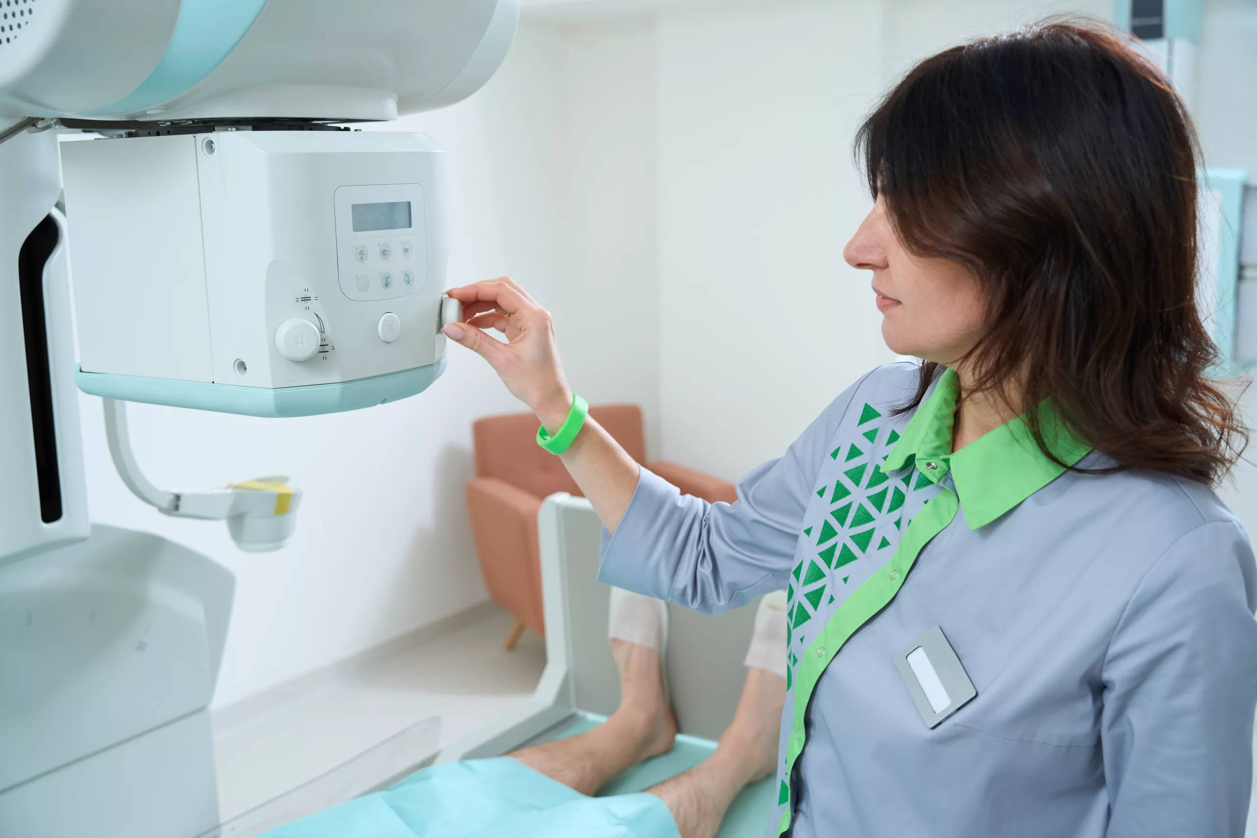
Procedure of ICGA
The ICGA procedure involves several steps:
- Patient Preparation: The patient is positioned comfortably, and the eye to be examined is prepared. Topical anesthetic drops may be applied to numb the eye and reduce discomfort.
- Injection of Dye: A small amount of indocyanine green dye is injected intravenously, usually into a vein in the arm. The dye quickly travels through the bloodstream and reaches the choroidal circulation.
- Image Acquisition: As the dye circulates, a specialized camera equipped with a near-infrared filter captures sequential images of the choroid. The images are taken at various time intervals to capture different phases of dye distribution.
- Post-Procedure Care: After the images are obtained, the patient may experience some temporary visual disturbances, but these typically resolve quickly. The eye is monitored for any adverse reactions to the dye.
Applications of ICGA
ICGA is used to diagnose and manage a range of ocular conditions, including:
- Age-Related Macular Degeneration (AMD): ICGA helps in identifying choroidal neovascularization (CNV), a common complication in AMD.
- Choroidal Neovascularization (CNV): ICGA provides detailed images of CNV, aiding in the assessment and treatment planning.
- Central Serous Chorioretinopathy (CSCR): ICGA can detect abnormal choroidal leakage and assess the extent of fluid accumulation.
- Polypoidal Choroidal Vasculopathy (PCV): ICGA helps in visualizing the characteristic polypoidal lesions associated with PCV.
- Choroidal Melanoma: ICGA assists in evaluating the extent and characteristics of choroidal tumors.
Advantages of ICGA
- Enhanced Choroidal Visualization: ICGA provides superior imaging of the choroid compared to other modalities, allowing for detailed assessment of choroidal pathology.
- Early Detection: The technique can detect abnormalities in the choroidal circulation before they become apparent in other imaging modalities.
- Dynamic Imaging: ICGA offers real-time imaging of the choroidal blood flow and leakage, aiding in dynamic assessment and treatment monitoring.
Limitations of ICGA
- Invasiveness: As with any procedure involving intravenous dye, there is a risk of allergic reactions or adverse effects, although these are rare.
- Cost: ICGA is more expensive than some other imaging techniques, which may limit its availability in certain settings.
- Limited Resolution: While ICGA provides excellent choroidal detail, it may not always be as effective in visualizing retinal structures compared to fluorescein angiography.
Recent Advancements in ICGA
Recent developments in ICGA technology have focused on improving image quality and expanding its clinical applications:
- Enhanced Imaging Systems: Newer ICGA systems offer higher resolution and faster image acquisition, providing more detailed and accurate choroidal images.
- Combination Techniques: Integrating ICGA with other imaging modalities, such as optical coherence tomography (OCT), enhances diagnostic capabilities and provides a comprehensive view of ocular conditions.
- Dye Formulations: Advances in dye formulations aim to reduce potential side effects and improve the safety profile of ICGA.
Conclusion
Indocyanine Green Angiography (ICGA) is a vital tool in modern ophthalmology, offering detailed insights into choroidal pathology that are crucial for diagnosing and managing various ocular conditions. Despite its limitations, the advantages of ICGA, including enhanced choroidal visualization and dynamic imaging capabilities, make it an indispensable part of the diagnostic and therapeutic arsenal in eye care. As technology continues to advance, ICGA will likely see further improvements, enhancing its effectiveness and expanding its applications in ocular health.
World Eye Care Foundation’s eyecare.live brings you the latest information from various industry sources and experts in eye health and vision care. Please consult with your eye care provider for more general information and specific eye conditions. We do not provide any medical advice, suggestions or recommendations in any health conditions.
Commonly Asked Questions
Patients should follow any specific instructions given by their healthcare provider. Generally, it is advised to avoid eating or drinking for a few hours before the procedure. Inform your provider of any allergies, especially to contrast dyes or medications. Wearing comfortable clothing and bringing someone to accompany you may also be helpful, as you might feel a bit disoriented after the procedure.
ICGA can be used in pediatric patients if necessary, but it is less commonly performed in children compared to adults. The decision to use ICGA in pediatric cases depends on the specific clinical situation and the child’s overall health. Special considerations and precautions are taken to ensure the safety and comfort of younger patients.
The frequency of ICGA depends on the specific condition being monitored and the recommendations of the ophthalmologist. For chronic conditions such as age-related macular degeneration or choroidal neovascularization, ICGA may be performed periodically to assess disease progression and response to treatment. Your healthcare provider will determine the appropriate interval based on your individual needs.
Yes, alternatives to ICGA include fluorescein angiography, optical coherence tomography (OCT), and OCT angiography. Each technique has its strengths and is used based on the specific needs of the diagnosis. For example, OCT is excellent for detailed cross-sectional images of the retina, while OCT angiography provides non-invasive imaging of blood flow.
Yes, ICGA is highly effective in detecting early-stage choroidal diseases. Its ability to provide detailed images of the choroidal blood vessels allows for early detection of abnormalities, such as choroidal neovascularization, that may not be visible with other imaging techniques.
Before the procedure, you may be asked to avoid eating for a few hours. During the procedure, you will be given an intravenous injection of indocyanine green dye, and the imaging will be conducted using a specialized camera. After the procedure, you might experience temporary visual changes or mild discomfort. It’s important to rest and avoid strenuous activities for a short period. Your doctor will provide specific instructions for post-procedure care.
The ICGA procedure typically takes about 30 to 60 minutes. This includes the time required for dye injection, image acquisition, and any necessary preparation or post-procedure monitoring. The actual imaging portion usually lasts just a few minutes.
ICGA is generally safe for most patients, but it may not be suitable for those with certain allergies, especially to iodides or contrast dyes, or those with severe kidney problems, as the dye is excreted through the kidneys. It is important for patients to inform their healthcare provider about any known allergies or medical conditions before undergoing the procedure.
ICGA and fluorescein angiography are both used to visualize blood vessels in the eye, but they differ in their target areas and imaging techniques. ICGA primarily highlights the choroidal vessels using indocyanine green dye and near-infrared light, offering detailed images of the choroid. In contrast, fluorescein angiography uses fluorescein dye and visible light to primarily visualize retinal blood vessels. ICGA is particularly useful for assessing choroidal conditions that fluorescein angiography may not fully reveal.
Common side effects of ICGA include temporary visual disturbances, such as changes in vision or seeing flashes of light. Some patients may also experience nausea or mild discomfort from the injection. Rarely, more serious side effects, such as allergic reactions to the dye, may occur. If any severe symptoms develop, it is important to contact a healthcare provider immediately.


news via inbox
Subscribe here to get latest updates !


