Welcome to World Eye Care Foundation! Today is : September 28, 2024

<div id="""" class="fusion-container-anchor"><div class="fusion-fullwidth fullwidth-box fusion-builder-row-4-1 fusion-flex-container video-background "" nonhundred-percent-fullwidth non-hundred-percent-height-scrolling fusion-no-small-visibility fusion-no-large-visibility lazyload" style="--awb-background-position:"center;--awb-background-repeat:"no-repeat";--awb-background-blend-mode:overlay;--awb-border-color:"";--awb-border-style:"solid";--awb-border-radius-top-left:0px;--awb-border-radius-top-right:0px;--awb-border-radius-bottom-right:0px;--awb-border-radius-bottom-left:0px;--awb-padding-top:;--awb-padding-right:;--awb-padding-bottom:;--awb-padding-left:;--awb-background-color:rgba(255,255,255,0.5);--awb-flex-wrap:wrap;" data-bg="""" id="""" ><div style='opacity:0;' class='fusion-background-video-wrapper' id='video-1' data-youtube-video-id='""' data-mute='"yes"' data-loop='0' data-loop-adjustment='' data-video-aspect-ratio='"16:9"'><div class='fusion-container-video-bg' id='video-2-inner'></div></div><div class="fullwidth-overlay" style="background-color:rgba(255,255,255,0.5);"></div><div class="fusion-builder-row fusion-row fusion-flex-align-items-flex-start fusion-flex-content-wrap" style="width:1302px !important;max-width:1302px !important;margin-left: calc(-5% / 2 );margin-right: calc(-5% / 2 );"><div class="fusion-layout-column fusion_builder_column fusion-builder-column-7 fusion_builder_column_"1_1" "1_1" fusion-flex-column "" fusion-no-small-visibility fusion-no-large-visibility fusion-column-inner-bg-wrapper fusion-animated" style="--awb-padding-top:;--awb-padding-right:;--awb-padding-bottom:;--awb-padding-left:;--awb-inner-bg-color:"";--awb-inner-bg-color-hover:"";--awb-inner-bg-position:"left;--awb-inner-bg-repeat:"no-repeat";--awb-border-color:"";--awb-border-top:;--awb-border-right:;--awb-border-bottom:;--awb-border-left:;--awb-border-style:"solid";--awb-margin-top-large:"0px";--awb-spacing-right-large:2.5%;--awb-margin-bottom-large:"0px";--awb-spacing-left-large:2.5%;--awb-spacing-right-medium:2.5%;--awb-spacing-left-medium:2.5%;--awb-width-small:100%;--awb-spacing-right-small:2.375%;--awb-spacing-left-small:2.375%;" data-animationType="""In"left"" data-animationDuration=""0.3"" data-animationOffset="top-into-view" id=""""><span class="fusion-column-inner-bg hover-type-"none""><a class="fusion-column-anchor" href=""""><span class="fusion-column-inner-bg-image lazyload" data-bg=""""></span></a></span><div class="fusion-column-wrapper fusion-flex-justify-content-flex-start fusion-content-layout-column fusion-column-has-bg-image" data-bg-url=""""><div class="fusion-title title fusion-title-1 fusion-no-small-visibility fusion-no-large-visibility fusion-sep-none fusion-title-"text" fusion-loop-"off" fusion-title-size-div fusion-animated fusion-has-text-overflow """ style="--awb-text-color:#ffffff;--awb-margin-top:;--awb-margin-right:;--awb-margin-bottom:;--awb-margin-left:;--awb-margin-top-small:;--awb-margin-right-small:;--awb-margin-bottom-small:;--awb-margin-left-small:;--awb-margin-top-medium:;--awb-margin-right-medium:;--awb-margin-bottom-medium:;--awb-margin-left-medium:;--awb-text-stroke-size:"1";--awb-text-stroke-color:#ffffff;--awb-sep-color:#ffffff;--awb-font-size:;--awb-text-overflow:"none";" data-animationType="""In"left"" data-animationDuration=""0.3"" data-animationOffset="""" id=""""><h"2" class="fusion-title-heading title-heading-"left" md-text-align-"" sm-text-align-""" style="font-family:"""";font-style:normal;font-weight:"";margin:0;font-size:1em;letter-spacing:;text-transform:"";line-height:;"><a href="""" target=""_self"">Introduction</a></h"2"></div><div class="fusion-text fusion-text-2 fusion-animated fusion-no-small-visibility fusion-no-large-visibility fusion-text-no-margin fusion-form-field-hidden """ style="--awb-content-alignment:"";--awb-font-size:;--awb-line-height:;--awb-letter-spacing:;--awb-text-transform:"";--awb-text-color:#ffffff;--awb-margin-top:;--awb-margin-right:;--awb-margin-bottom:;--awb-margin-left:;--awb-text-font-family:"""";--awb-text-font-style:normal;--awb-text-font-weight:"";" data-animationType="""In"left"" data-animationDuration=""0.3"" data-animationOffset="""" data-form-element-name="fusion_text_2" id=""""><p> Myopic macular degeneration is a complex eye condition that primarily affects individuals with severe nearsightedness (myopia). In this comprehensive guide, we will delve into the intricacies of myopic macular degeneration, exploring its causes, symptoms, diagnosis, and available treatment options. By shedding light on this condition, we aim to empower readers with valuable insights into managing and understanding myopic macular degeneration effectively. </p>
</div><div class="fusion-title title fusion-title-2 fusion-no-small-visibility fusion-no-large-visibility fusion-sep-none fusion-title-"text" fusion-loop-"off" fusion-title-size-div fusion-animated fusion-has-text-overflow """ style="--awb-text-color:#ffffff;--awb-margin-top:;--awb-margin-right:;--awb-margin-bottom:;--awb-margin-left:;--awb-margin-top-small:;--awb-margin-right-small:;--awb-margin-bottom-small:;--awb-margin-left-small:;--awb-margin-top-medium:;--awb-margin-right-medium:;--awb-margin-bottom-medium:;--awb-margin-left-medium:;--awb-text-stroke-size:"1";--awb-text-stroke-color:#ffffff;--awb-sep-color:#ffffff;--awb-font-size:;--awb-text-overflow:"none";" data-animationType="""In"left"" data-animationDuration=""0.3"" data-animationOffset="""" id=""""><h"2" class="fusion-title-heading title-heading-"left" md-text-align-"" sm-text-align-""" style="font-family:"""";font-style:normal;font-weight:"";margin:0;font-size:1em;letter-spacing:;text-transform:"";line-height:;"><a href="""" target=""_self"">Understanding Myopic Macular Degeneration</a></h"2"></div><div class="fusion-text fusion-text-3 fusion-animated fusion-no-small-visibility fusion-no-large-visibility fusion-text-no-margin fusion-form-field-hidden """ style="--awb-content-alignment:"";--awb-font-size:;--awb-line-height:;--awb-letter-spacing:;--awb-text-transform:"";--awb-text-color:#ffffff;--awb-margin-top:;--awb-margin-right:;--awb-margin-bottom:;--awb-margin-left:;--awb-text-font-family:"""";--awb-text-font-style:normal;--awb-text-font-weight:"";" data-animationType="""In"left"" data-animationDuration=""0.3"" data-animationOffset="""" data-form-element-name="fusion_text_3" id=""""><p> Myopic macular degeneration is a subtype of macular degeneration, a progressive eye condition characterized by the deterioration of the macula—the central part of the retina responsible for sharp, central vision. Unlike age-related macular degeneration (AMD), which typically affects older adults, myopic macular degeneration primarily occurs in individuals with high degrees of myopia (nearsightedness). High myopia is often defined as a refractive error greater than -6.00 diopters. In severe myopia, the eyeball becomes elongated, leading to structural changes in the retina, particularly in the macula. These changes may include thinning of the macular tissue, stretching of the retinal layers, and alterations in the blood supply to the macula. Over time, these structural abnormalities can contribute to vision loss and impairment. </p>
</div><div class="fusion-title title fusion-title-3 fusion-no-small-visibility fusion-no-large-visibility fusion-sep-none fusion-title-"text" fusion-loop-"off" fusion-title-size-div fusion-animated fusion-has-text-overflow """ style="--awb-text-color:#ffffff;--awb-margin-top:;--awb-margin-right:;--awb-margin-bottom:;--awb-margin-left:;--awb-margin-top-small:;--awb-margin-right-small:;--awb-margin-bottom-small:;--awb-margin-left-small:;--awb-margin-top-medium:;--awb-margin-right-medium:;--awb-margin-bottom-medium:;--awb-margin-left-medium:;--awb-text-stroke-size:"1";--awb-text-stroke-color:#ffffff;--awb-sep-color:#ffffff;--awb-font-size:;--awb-text-overflow:"none";" data-animationType="""In"left"" data-animationDuration=""0.3"" data-animationOffset="""" id=""""><h"2" class="fusion-title-heading title-heading-"left" md-text-align-"" sm-text-align-""" style="font-family:"""";font-style:normal;font-weight:"";margin:0;font-size:1em;letter-spacing:;text-transform:"";line-height:;"><a href="""" target=""_self"">Causes of Myopic Macular Degeneration</a></h"2"></div><div class="fusion-text fusion-text-4 fusion-animated fusion-no-small-visibility fusion-no-large-visibility fusion-text-no-margin fusion-form-field-hidden """ style="--awb-content-alignment:"";--awb-font-size:;--awb-line-height:;--awb-letter-spacing:;--awb-text-transform:"";--awb-text-color:#ffffff;--awb-margin-top:;--awb-margin-right:;--awb-margin-bottom:;--awb-margin-left:;--awb-text-font-family:"""";--awb-text-font-style:normal;--awb-text-font-weight:"";" data-animationType="""In"left"" data-animationDuration=""0.3"" data-animationOffset="""" data-form-element-name="fusion_text_4" id=""""><p> Several factors contribute to the development and progression of myopic macular degeneration: High Myopia: Individuals with severe nearsightedness are at higher risk of developing myopic macular degeneration due to the elongation of the eyeball. The greater the degree of myopia, the higher the risk of associated ocular complications. Axial Length Elongation: High myopia is associated with excessive elongation of the eyeball, known as axial length elongation. This elongation can lead to structural changes in the retina, including thinning and stretching of the macula, making it more susceptible to degenerative changes. Genetic Predisposition: There may be a genetic component to myopic macular degeneration, with certain gene variations increasing an individual's susceptibility to the condition. Family history of myopia or macular degeneration may also play a role in predisposing individuals to this condition. Environmental Factors: Environmental factors, such as prolonged near work, limited outdoor activities, and improper visual hygiene during childhood and adolescence, can contribute to the progression of myopia and subsequent macular changes. Adequate exposure to natural light and maintaining proper visual habits may help mitigate these risk factors. </p>
</div><div class="fusion-title title fusion-title-4 fusion-no-small-visibility fusion-no-large-visibility fusion-sep-none fusion-title-"text" fusion-loop-"off" fusion-title-size-div fusion-animated fusion-has-text-overflow """ style="--awb-text-color:#ffffff;--awb-margin-top:;--awb-margin-right:;--awb-margin-bottom:;--awb-margin-left:;--awb-margin-top-small:;--awb-margin-right-small:;--awb-margin-bottom-small:;--awb-margin-left-small:;--awb-margin-top-medium:;--awb-margin-right-medium:;--awb-margin-bottom-medium:;--awb-margin-left-medium:;--awb-text-stroke-size:"1";--awb-text-stroke-color:#ffffff;--awb-sep-color:#ffffff;--awb-font-size:;--awb-text-overflow:"none";" data-animationType="""In"left"" data-animationDuration=""0.3"" data-animationOffset="""" id=""""><h"2" class="fusion-title-heading title-heading-"left" md-text-align-"" sm-text-align-""" style="font-family:"""";font-style:normal;font-weight:"";margin:0;font-size:1em;letter-spacing:;text-transform:"";line-height:;"><a href="""" target=""_self"">Symptoms of Myopic Macular Degeneration</a></h"2"></div><div class="fusion-text fusion-text-5 fusion-animated fusion-no-small-visibility fusion-no-large-visibility fusion-text-no-margin fusion-form-field-hidden """ style="--awb-content-alignment:"";--awb-font-size:;--awb-line-height:;--awb-letter-spacing:;--awb-text-transform:"";--awb-text-color:#ffffff;--awb-margin-top:;--awb-margin-right:;--awb-margin-bottom:;--awb-margin-left:;--awb-text-font-family:"""";--awb-text-font-style:normal;--awb-text-font-weight:"";" data-animationType="""In"left"" data-animationDuration=""0.3"" data-animationOffset="""" data-form-element-name="fusion_text_5" id=""""><p> The symptoms of myopic macular degeneration may vary depending on the severity and progression of the condition: Blurred Central Vision: Individuals may experience difficulty seeing objects clearly, especially when looking at things up close or reading fine print. Central Scotomas: Dark or empty areas may appear in the central vision field, affecting the ability to perceive details and colors. Metamorphopsia: Straight lines may appear distorted or wavy, indicating changes in the macular structure. Difficulty in Low Light: Reduced ability to see clearly in dimly lit environments, as the degenerative changes in the macula may impact visual sensitivity. It's essential to note that individuals with myopic macular degeneration may not experience symptoms in the early stages of the condition. Regular eye examinations are crucial for early detection and management. </p>
</div><div class="fusion-title title fusion-title-5 fusion-no-small-visibility fusion-no-large-visibility fusion-sep-none fusion-title-"text" fusion-loop-"off" fusion-title-size-div fusion-animated fusion-has-text-overflow """ style="--awb-text-color:#ffffff;--awb-margin-top:;--awb-margin-right:;--awb-margin-bottom:;--awb-margin-left:;--awb-margin-top-small:;--awb-margin-right-small:;--awb-margin-bottom-small:;--awb-margin-left-small:;--awb-margin-top-medium:;--awb-margin-right-medium:;--awb-margin-bottom-medium:;--awb-margin-left-medium:;--awb-text-stroke-size:"1";--awb-text-stroke-color:#ffffff;--awb-sep-color:#ffffff;--awb-font-size:;--awb-text-overflow:"none";" data-animationType="""In"left"" data-animationDuration=""0.3"" data-animationOffset="""" id=""""><h"2" class="fusion-title-heading title-heading-"left" md-text-align-"" sm-text-align-""" style="font-family:"""";font-style:normal;font-weight:"";margin:0;font-size:1em;letter-spacing:;text-transform:"";line-height:;"><a href="""" target=""_self"">Diagnosis and Evaluation</a></h"2"></div><div class="fusion-text fusion-text-6 fusion-animated fusion-no-small-visibility fusion-no-large-visibility fusion-text-no-margin fusion-form-field-hidden """ style="--awb-content-alignment:"";--awb-font-size:;--awb-line-height:;--awb-letter-spacing:;--awb-text-transform:"";--awb-text-color:#ffffff;--awb-margin-top:;--awb-margin-right:;--awb-margin-bottom:;--awb-margin-left:;--awb-text-font-family:"""";--awb-text-font-style:normal;--awb-text-font-weight:"";" data-animationType="""In"left"" data-animationDuration=""0.3"" data-animationOffset="""" data-form-element-name="fusion_text_6" id=""""><p> Diagnosing myopic macular degeneration involves a comprehensive eye examination conducted by an eye care professional, which may include: Visual Acuity Test: Assessing the clarity and sharpness of vision using an eye chart. Dilated Fundus Examination: Examining the back of the eye, including the macula, using specialized instruments to evaluate for structural abnormalities and signs of degeneration. Optical Coherence Tomography (OCT): Producing high-resolution cross-sectional images of the retina, allowing for detailed assessment of macular thickness, integrity, and any abnormalities. Fluorescein Angiography: Evaluating the retinal blood vessels and identifying any leakage, blockages, or abnormalities using a fluorescent dye and specialized imaging techniques. Additional tests, such as fundus autofluorescence and electroretinography, may also be performed to assess the extent and severity of macular degeneration. </p>
</div><div class="fusion-title title fusion-title-6 fusion-no-small-visibility fusion-no-large-visibility fusion-sep-none fusion-title-"text" fusion-loop-"off" fusion-title-size-div fusion-animated fusion-has-text-overflow """ style="--awb-text-color:#ffffff;--awb-margin-top:;--awb-margin-right:;--awb-margin-bottom:;--awb-margin-left:;--awb-margin-top-small:;--awb-margin-right-small:;--awb-margin-bottom-small:;--awb-margin-left-small:;--awb-margin-top-medium:;--awb-margin-right-medium:;--awb-margin-bottom-medium:;--awb-margin-left-medium:;--awb-text-stroke-size:"1";--awb-text-stroke-color:#ffffff;--awb-sep-color:#ffffff;--awb-font-size:;--awb-text-overflow:"none";" data-animationType="""In"left"" data-animationDuration=""0.3"" data-animationOffset="""" id=""""><h"2" class="fusion-title-heading title-heading-"left" md-text-align-"" sm-text-align-""" style="font-family:"""";font-style:normal;font-weight:"";margin:0;font-size:1em;letter-spacing:;text-transform:"";line-height:;"><a href="""" target=""_self"">Treatment Options</a></h"2"></div><div class="fusion-text fusion-text-7 fusion-animated fusion-no-small-visibility fusion-no-large-visibility fusion-text-no-margin fusion-form-field-hidden """ style="--awb-content-alignment:"";--awb-font-size:;--awb-line-height:;--awb-letter-spacing:;--awb-text-transform:"";--awb-text-color:#ffffff;--awb-margin-top:;--awb-margin-right:;--awb-margin-bottom:;--awb-margin-left:;--awb-text-font-family:"""";--awb-text-font-style:normal;--awb-text-font-weight:"";" data-animationType="""In"left"" data-animationDuration=""0.3"" data-animationOffset="""" data-form-element-name="fusion_text_7" id=""""><p> While there is currently no cure for myopic macular degeneration, several treatment modalities aim to manage the condition, alleviate symptoms, and preserve vision: Lifestyle Modifications: Lifestyle changes and behavioral interventions may help slow the progression of myopic macular degeneration and reduce the risk of complications. These may include: Control of Myopia Progression: Implementing strategies such as orthokeratology (corneal reshaping therapy), multifocal contact lenses, and atropine eye drops to slow the progression of myopia and reduce axial length elongation. Healthy Habits: Encouraging outdoor activities, limiting screen time, maintaining proper posture, and practicing good visual hygiene to promote ocular health and reduce eye strain. Intravitreal Injections: Intravitreal injections of anti-vascular endothelial growth factor (anti-VEGF) agents may be administered to reduce abnormal blood vessel growth and leakage in the macula, thereby slowing the progression of myopic macular degeneration and preserving vision. Photodynamic Therapy (PDT): Photodynamic therapy involves the administration of a light-sensitive drug followed by targeted laser treatment to selectively destroy abnormal blood vessels in the macula while minimizing damage to healthy tissue. PDT may be used to treat certain cases of myopic macular degeneration associated with choroidal neovascularization. Vitrectomy: In severe cases of myopic macular degeneration with significant vitreoretinal traction or hemorrhage, vitrectomy surgery may be considered. Vitrectomy involves the removal of the vitreous gel and any associated scar tissue from the eye, thereby relieving traction and improving visual outcomes. The choice of treatment modality depends on various factors, including the severity of macular degeneration, the presence of complications, and individual patient characteristics. A comprehensive evaluation by an eye care professional is essential to determine the most appropriate treatment approach. </p>
</div><div class="fusion-title title fusion-title-7 fusion-no-small-visibility fusion-no-large-visibility fusion-sep-none fusion-title-"text" fusion-loop-"off" fusion-title-size-div fusion-animated fusion-has-text-overflow """ style="--awb-text-color:#ffffff;--awb-margin-top:;--awb-margin-right:;--awb-margin-bottom:;--awb-margin-left:;--awb-margin-top-small:;--awb-margin-right-small:;--awb-margin-bottom-small:;--awb-margin-left-small:;--awb-margin-top-medium:;--awb-margin-right-medium:;--awb-margin-bottom-medium:;--awb-margin-left-medium:;--awb-text-stroke-size:"1";--awb-text-stroke-color:#ffffff;--awb-sep-color:#ffffff;--awb-font-size:;--awb-text-overflow:"none";" data-animationType="""In"left"" data-animationDuration=""0.3"" data-animationOffset="""" id=""""><h"2" class="fusion-title-heading title-heading-"left" md-text-align-"" sm-text-align-""" style="font-family:"""";font-style:normal;font-weight:"";margin:0;font-size:1em;letter-spacing:;text-transform:"";line-height:;"><a href="""" target=""_self"">Prevention Strategies</a></h"2"></div><div class="fusion-text fusion-text-8 fusion-animated fusion-no-small-visibility fusion-no-large-visibility fusion-text-no-margin fusion-form-field-hidden """ style="--awb-content-alignment:"";--awb-font-size:;--awb-line-height:;--awb-letter-spacing:;--awb-text-transform:"";--awb-text-color:#ffffff;--awb-margin-top:;--awb-margin-right:;--awb-margin-bottom:;--awb-margin-left:;--awb-text-font-family:"""";--awb-text-font-style:normal;--awb-text-font-weight:"";" data-animationType="""In"left"" data-animationDuration=""0.3"" data-animationOffset="""" data-form-element-name="fusion_text_8" id=""""><p> Preventing the progression of myopic macular degeneration involves a multifaceted approach aimed at slowing the progression of myopia, promoting ocular health, and reducing modifiable risk factors. Key prevention strategies include: Regular Eye Examinations: Undergoing comprehensive eye examinations at regular intervals to monitor changes in vision, assess macular health, and detect early signs of macular degeneration. Myopia Control Measures: Implementing strategies to slow the progression of myopia in children and adolescents, such as orthokeratology, multifocal contact lenses, atropine eye drops, and behavioral modifications. Lifestyle Modifications: Encouraging healthy habits, including outdoor activities, balanced nutrition, adequate hydration, proper posture, and regular breaks from near work to reduce eye strain and promote ocular health. Visual Hygiene Practices: Practicing good visual hygiene, such as maintaining proper lighting, reducing screen time, taking regular breaks during prolonged near work, and ensuring optimal ergonomic conditions to minimize visual stress and discomfort. Environmental Interventions: Promoting initiatives to create myopia-friendly environments, such as implementing outdoor learning programs in schools, optimizing classroom lighting, and raising awareness about the importance of outdoor activities for ocular health. It's important for individuals with myopia and those at risk of myopic macular degeneration to proactively engage in preventive measures and adopt a holistic approach to eye care to preserve vision and promote lifelong ocular health. </p>
</div><div class="fusion-title title fusion-title-8 fusion-no-small-visibility fusion-no-large-visibility fusion-sep-none fusion-title-"text" fusion-loop-"off" fusion-title-size-div fusion-animated fusion-has-text-overflow """ style="--awb-text-color:#ffffff;--awb-margin-top:;--awb-margin-right:;--awb-margin-bottom:;--awb-margin-left:;--awb-margin-top-small:;--awb-margin-right-small:;--awb-margin-bottom-small:;--awb-margin-left-small:;--awb-margin-top-medium:;--awb-margin-right-medium:;--awb-margin-bottom-medium:;--awb-margin-left-medium:;--awb-text-stroke-size:"1";--awb-text-stroke-color:#ffffff;--awb-sep-color:#ffffff;--awb-font-size:;--awb-text-overflow:"none";" data-animationType="""In"left"" data-animationDuration=""0.3"" data-animationOffset="""" id=""""><h"2" class="fusion-title-heading title-heading-"left" md-text-align-"" sm-text-align-""" style="font-family:"""";font-style:normal;font-weight:"";margin:0;font-size:1em;letter-spacing:;text-transform:"";line-height:;"><a href="""" target=""_self"">Conclusion</a></h"2"></div><div class="fusion-text fusion-text-9 fusion-animated fusion-no-small-visibility fusion-no-large-visibility fusion-text-no-margin fusion-form-field-hidden """ style="--awb-content-alignment:"";--awb-font-size:;--awb-line-height:;--awb-letter-spacing:;--awb-text-transform:"";--awb-text-color:#ffffff;--awb-margin-top:;--awb-margin-right:;--awb-margin-bottom:;--awb-margin-left:;--awb-text-font-family:"""";--awb-text-font-style:normal;--awb-text-font-weight:"";" data-animationType="""In"left"" data-animationDuration=""0.3"" data-animationOffset="""" data-form-element-name="fusion_text_9" id=""""><p> Myopic macular degeneration poses significant challenges for individuals with severe nearsightedness, affecting central vision and quality of life. By understanding the causes, symptoms, and available treatment options, individuals can take proactive steps to manage the condition effectively. Regular eye examinations, lifestyle modifications, and early intervention are essential for preserving vision and minimizing the impact of myopic macular degeneration on ocular health. If you or someone you know is experiencing symptoms of myopic macular degeneration, consult an eye care professional for personalized evaluation and management strategies. </p>
</div><div class="fusion-text fusion-text-10 fusion-animated fusion-no-small-visibility fusion-no-large-visibility fusion-text-no-margin fusion-form-field-hidden """ style="--awb-content-alignment:"";--awb-font-size:"var(--awb-typography5-font-size)";--awb-line-height:;--awb-letter-spacing:;--awb-text-transform:"";--awb-text-color:#ffffff;--awb-margin-top:;--awb-margin-right:;--awb-margin-bottom:;--awb-margin-left:;--awb-text-font-family:"""";--awb-text-font-style:normal;--awb-text-font-weight:"";" data-animationType="""In"left"" data-animationDuration=""0.3"" data-animationOffset="""" data-form-element-name="fusion_text_10" id=""""><p> World Eye Care Foundation’s eyecare.live brings you the latest information from various industry sources and experts in eye health and vision care. Please consult with your eye care provider for more general information and specific eye conditions. We do not provide any medical advice, suggestions or recommendations in any health conditions. </p>
</div></div></div></div></div></div>

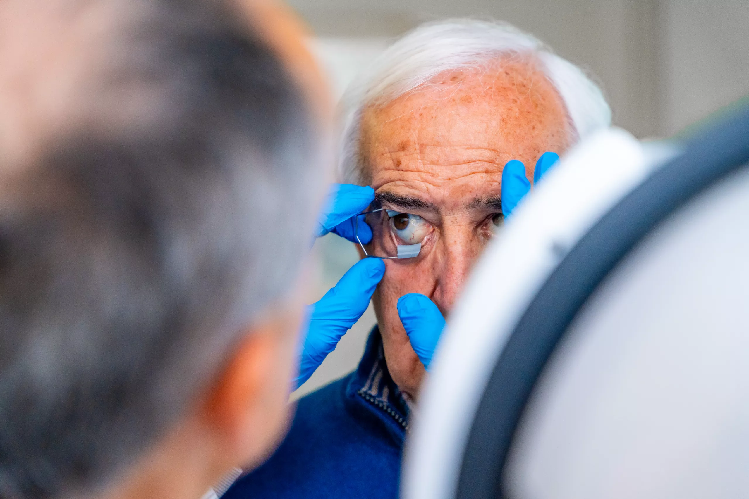
Photo credit to Andri Munazir

Demystifying Myopic Macular Degeneration

Introduction

Myopic macular degeneration is a complex eye condition that primarily affects individuals with severe nearsightedness (myopia). In this comprehensive guide, we will delve into the intricacies of myopic macular degeneration, exploring its causes, symptoms, diagnosis, and available treatment options. By shedding light on this condition, we aim to empower readers with valuable insights into managing and understanding myopic macular degeneration effectively.

Understanding Myopic Macular Degeneration

Myopic macular degeneration is a subtype of macular degeneration, a progressive eye condition characterized by the deterioration of the macula—the central part of the retina responsible for sharp, central vision. Unlike age-related macular degeneration (AMD), which typically affects older adults, myopic macular degeneration primarily occurs in individuals with high degrees of myopia (nearsightedness). High myopia is often defined as a refractive error greater than -6.00 diopters.

In severe myopia, the eyeball becomes elongated, leading to structural changes in the retina, particularly in the macula. These changes may include thinning of the macular tissue, stretching of the retinal layers, and alterations in the blood supply to the macula. Over time, these structural abnormalities can contribute to vision loss and impairment.

Causes of Myopic Macular Degeneration

Several factors contribute to the development and progression of myopic macular degeneration:

  1. High Myopia: Individuals with severe nearsightedness are at higher risk of developing myopic macular degeneration due to the elongation of the eyeball. The greater the degree of myopia, the higher the risk of associated ocular complications.
  2. Axial Length Elongation: High myopia is associated with excessive elongation of the eyeball, known as axial length elongation. This elongation can lead to structural changes in the retina, including thinning and stretching of the macula, making it more susceptible to degenerative changes.
  3. Genetic Predisposition: There may be a genetic component to myopic macular degeneration, with certain gene variations increasing an individual’s susceptibility to the condition. Family history of myopia or macular degeneration may also play a role in predisposing individuals to this condition.
  4. Environmental Factors: Environmental factors, such as prolonged near work, limited outdoor activities, and improper visual hygiene during childhood and adolescence, can contribute to the progression of myopia and subsequent macular changes. Adequate exposure to natural light and maintaining proper visual habits may help mitigate these risk factors.

Symptoms of Myopic Macular Degeneration

The symptoms of myopic macular degeneration may vary depending on the severity and progression of the condition:

  • Blurred Central Vision: Individuals may experience difficulty seeing objects clearly, especially when looking at things up close or reading fine print.
  • Central Scotomas: Dark or empty areas may appear in the central vision field, affecting the ability to perceive details and colors.
  • Metamorphopsia: Straight lines may appear distorted or wavy, indicating changes in the macular structure.
  • Difficulty in Low Light: Reduced ability to see clearly in dimly lit environments, as the degenerative changes in the macula may impact visual sensitivity.

It’s essential to note that individuals with myopic macular degeneration may not experience symptoms in the early stages of the condition. Regular eye examinations are crucial for early detection and management.

Diagnosis and Evaluation

Diagnosing myopic macular degeneration involves a comprehensive eye examination conducted by an eye care professional, which may include:

  • Visual Acuity Test: Assessing the clarity and sharpness of vision using an eye chart.
  • Dilated Fundus Examination: Examining the back of the eye, including the macula, using specialized instruments to evaluate for structural abnormalities and signs of degeneration.
  • Optical Coherence Tomography (OCT): Producing high-resolution cross-sectional images of the retina, allowing for detailed assessment of macular thickness, integrity, and any abnormalities.
  • Fluorescein Angiography: Evaluating the retinal blood vessels and identifying any leakage, blockages, or abnormalities using a fluorescent dye and specialized imaging techniques.

Additional tests, such as fundus autofluorescence and electroretinography, may also be performed to assess the extent and severity of macular degeneration.

Treatment Options

While there is currently no cure for myopic macular degeneration, several treatment modalities aim to manage the condition, alleviate symptoms, and preserve vision:

  1. Lifestyle Modifications: Lifestyle changes and behavioral interventions may help slow the progression of myopic macular degeneration and reduce the risk of complications. These may include:
    • Control of Myopia Progression: Implementing strategies such as orthokeratology (corneal reshaping therapy), multifocal contact lenses, and atropine eye drops to slow the progression of myopia and reduce axial length elongation.
    • Healthy Habits: Encouraging outdoor activities, limiting screen time, maintaining proper posture, and practicing good visual hygiene to promote ocular health and reduce eye strain.
  2. Intravitreal Injections: Intravitreal injections of anti-vascular endothelial growth factor (anti-VEGF) agents may be administered to reduce abnormal blood vessel growth and leakage in the macula, thereby slowing the progression of myopic macular degeneration and preserving vision.
  3. Photodynamic Therapy (PDT): Photodynamic therapy involves the administration of a light-sensitive drug followed by targeted laser treatment to selectively destroy abnormal blood vessels in the macula while minimizing damage to healthy tissue. PDT may be used to treat certain cases of myopic macular degeneration associated with choroidal neovascularization.
  4. Vitrectomy: In severe cases of myopic macular degeneration with significant vitreoretinal traction or hemorrhage, vitrectomy surgery may be considered. Vitrectomy involves the removal of the vitreous gel and any associated scar tissue from the eye, thereby relieving traction and improving visual outcomes.

The choice of treatment modality depends on various factors, including the severity of macular degeneration, the presence of complications, and individual patient characteristics. A comprehensive evaluation by an eye care professional is essential to determine the most appropriate treatment approach.

Prevention Strategies

Preventing the progression of myopic macular degeneration involves a multifaceted approach aimed at slowing the progression of myopia, promoting ocular health, and reducing modifiable risk factors. Key prevention strategies include:

  • Regular Eye Examinations: Undergoing comprehensive eye examinations at regular intervals to monitor changes in vision, assess macular health, and detect early signs of macular degeneration.
  • Myopia Control Measures: Implementing strategies to slow the progression of myopia in children and adolescents, such as orthokeratology, multifocal contact lenses, atropine eye drops, and behavioral modifications.
  • Lifestyle Modifications: Encouraging healthy habits, including outdoor activities, balanced nutrition, adequate hydration, proper posture, and regular breaks from near work to reduce eye strain and promote ocular health.
  • Visual Hygiene Practices: Practicing good visual hygiene, such as maintaining proper lighting, reducing screen time, taking regular breaks during prolonged near work, and ensuring optimal ergonomic conditions to minimize visual stress and discomfort.
  • Environmental Interventions: Promoting initiatives to create myopia-friendly environments, such as implementing outdoor learning programs in schools, optimizing classroom lighting, and raising awareness about the importance of outdoor activities for ocular health.

It’s important for individuals with myopia and those at risk of myopic macular degeneration to proactively engage in preventive measures and adopt a holistic approach to eye care to preserve vision and promote lifelong ocular health.

Conclusion

Myopic macular degeneration poses significant challenges for individuals with severe nearsightedness, affecting central vision and quality of life. By understanding the causes, symptoms, and available treatment options, individuals can take proactive steps to manage the condition effectively. Regular eye examinations, lifestyle modifications, and early intervention are essential for preserving vision and minimizing the impact of myopic macular degeneration on ocular health. If you or someone you know is experiencing symptoms of myopic macular degeneration, consult an eye care professional for personalized evaluation and management strategies.

World Eye Care Foundation’s eyecare.live brings you the latest information from various industry sources and experts in eye health and vision care. Please consult with your eye care provider for more general information and specific eye conditions. We do not provide any medical advice, suggestions or recommendations in any health conditions.

Commonly Asked Questions

Why is early detection important for myopic macular degeneration?2024-06-17T04:51:47+00:00

Early detection allows for timely management and intervention, potentially slowing down the progression of the condition and preserving vision.

What are the key prevention strategies for myopic macular degeneration?2024-06-17T04:51:14+00:00

Prevention strategies include regular eye exams, myopia control measures, healthy lifestyle habits, visual hygiene practices, and creating myopia-friendly environments.

What lifestyle modifications can help manage myopic macular degeneration?2024-06-17T04:51:00+00:00

Lifestyle changes such as controlling myopia progression, outdoor activities, limiting screen time, and maintaining proper posture can help manage the condition.

Can myopic macular degeneration be prevented?2024-06-17T04:50:45+00:00

Prevention strategies include regular eye examinations, myopia control measures, lifestyle modifications, visual hygiene practices, and environmental interventions.

What treatment options are available for myopic macular degeneration?2024-06-17T04:50:21+00:00

Treatment options include lifestyle modifications, intravitreal injections, photodynamic therapy (PDT), and vitrectomy surgery depending on the severity of the condition.

How is myopic macular degeneration diagnosed?2024-06-17T04:50:04+00:00

Diagnosis involves comprehensive eye examinations including visual acuity tests, dilated fundus examination, optical coherence tomography (OCT), and fluorescein angiography.

What are the causes of myopic macular degeneration apart from high myopia?2024-06-17T04:49:49+00:00

Apart from high myopia, causes may include axial length elongation, genetic predisposition, and environmental factors such as prolonged near work and limited outdoor activities.

What are the common symptoms of myopic macular degeneration?2024-06-17T04:49:33+00:00

Symptoms may include blurred central vision, central scotomas, metamorphopsia, and difficulty seeing in low light environments.

How does myopic macular degeneration differ from age-related macular degeneration (AMD)?2024-06-17T04:49:02+00:00

Myopic macular degeneration primarily affects individuals with high myopia, whereas AMD typically affects older adults. Myopic macular degeneration involves elongation of the eyeball and structural changes in the macula.

What is myopic macular degeneration?2024-06-17T04:48:36+00:00

Myopic macular degeneration is a subtype of macular degeneration primarily affecting individuals with severe nearsightedness (myopia). It involves structural changes in the macula, leading to vision impairment.

news via inbox

Subscribe here to get latest updates !

Go to Top