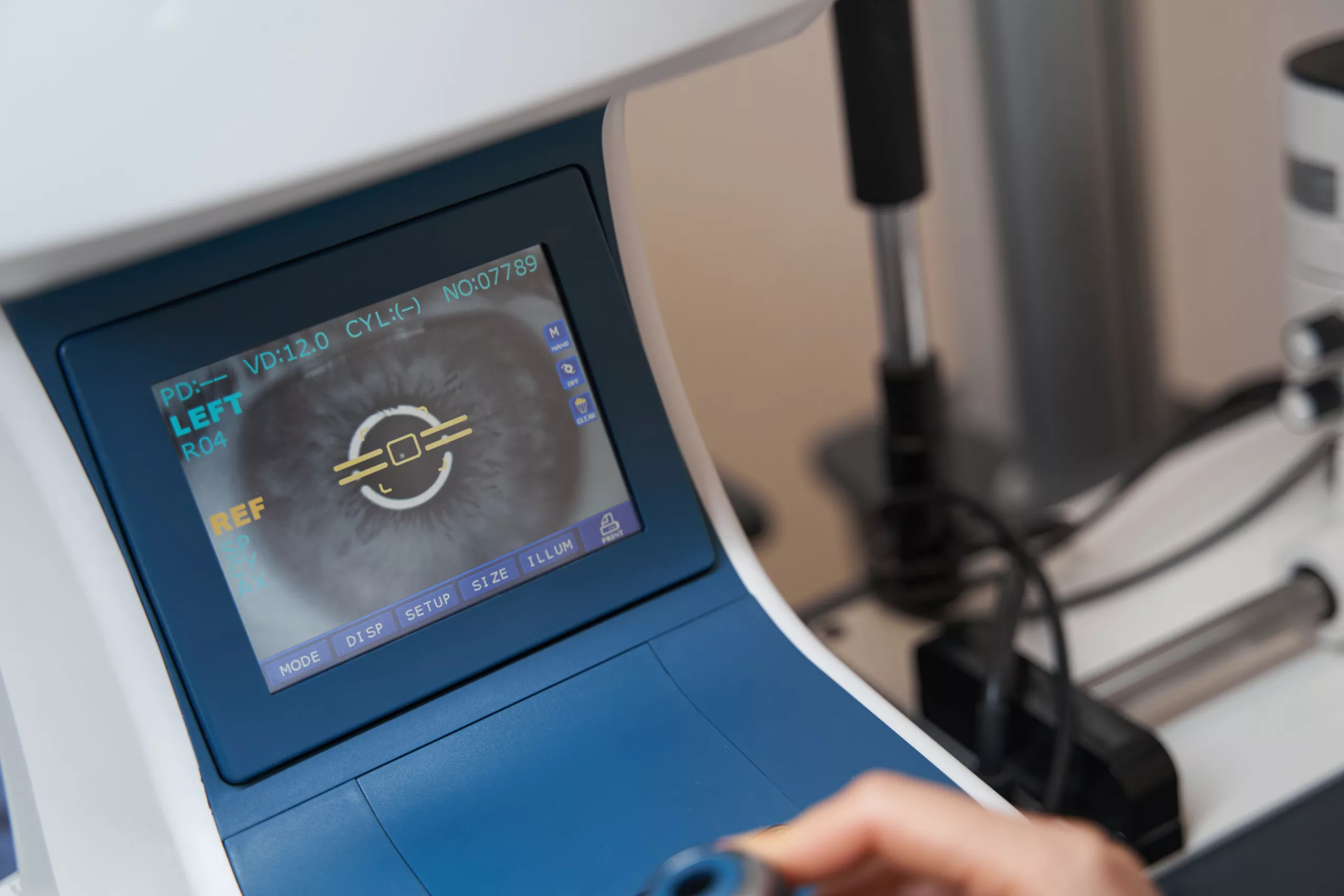
Goldmann Kinetic Perimetry: An In-Depth Exploration
Introduction
Goldmann Kinetic Perimetry (GKP) is a comprehensive method for evaluating the visual field, essential for diagnosing and managing various ocular and neurological conditions. Here’s a detailed examination of GKP, covering its principles, methodology, applications, benefits, limitations, and future directions, with additional insights.
What is Goldmann Kinetic Perimetry?
Goldmann Kinetic Perimetry is a visual field test that evaluates the range and sensitivity of a patient’s vision by moving a light stimulus across their visual field. Unlike static perimetry, which assesses fixed points of vision, GKP involves dynamic testing where the stimulus moves and the patient indicates when it is first perceived.
- Historical Context: Developed in the 1950s by Hans Goldmann, this method has been a cornerstone in visual field assessment due to its precision and adaptability. It is still widely used today for its effectiveness in detecting subtle visual field defects.
Principles of Goldmann Kinetic Perimetry
- Stimulus Size and Intensity:
- Stimulus Sizes: Goldmann perimetry uses five standard stimulus sizes, from I (smallest) to V (largest).
- Size I: Used for testing central vision.
- Size II: For testing central and mid-peripheral vision.
- Size III: For assessing mid-peripheral and peripheral vision.
- Size IV: For peripheral vision.
- Size V: For detecting very large visual field defects.
- Intensity Levels: Stimuli are presented at various intensities, often calibrated according to a standard brightness scale. For instance, a stimulus might be set to a level that corresponds to a specific number of decibels (dB) of light intensity.
- Stimulus Sizes: Goldmann perimetry uses five standard stimulus sizes, from I (smallest) to V (largest).
- Movement of Stimulus:
- Testing Procedure: The stimulus is moved in a predefined pattern (e.g., concentric circles or radial lines) from outside the visual field toward the center. The rate of movement and the distance between successive positions are adjusted based on the clinical need.
- Dynamic Testing: By tracking the patient’s responses to a moving stimulus, GKP can reveal scotomas (blind spots) and other visual field anomalies.
- Visual Field Mapping:
- Mapping Process: Responses from different regions are recorded and plotted on a visual field chart. This map shows areas where the patient’s vision is impaired, which helps in diagnosing conditions affecting the visual pathways.
- Data Representation: The results are often displayed as a graphical plot where the visual field is depicted with concentric circles, indicating different degrees of visual sensitivity.
Methodology
- Preparation:
- Patient Positioning: The patient sits in a darkened room in front of a perimeter machine. The head is placed on a chin rest and forehead bar to ensure stability.
- Calibration: The perimeter machine is calibrated according to the specific test parameters, including stimulus size and intensity. Calibration ensures accurate and reliable results.
- Test Execution:
- Stimulus Selection: The examiner chooses the appropriate stimulus size and intensity based on the patient’s condition. For instance, smaller stimuli might be used to test central vision in patients with suspected macular issues.
- Stimulus Movement: The stimulus is moved according to the selected testing pattern. The movement is usually slow and deliberate to allow the patient ample time to detect the stimulus.
- Patient Response:
- Detection: The patient indicates when they first see the stimulus by pressing a button or responding verbally. Consistent responses are crucial for accurate results.
- Response Accuracy: The test requires the patient’s full attention and cooperation. Distractions or non-cooperative behavior can affect the accuracy of the results.
- Data Collection:
- Recording Responses: The perimeter machine records the points where the stimulus is detected. This data is used to generate a visual field map.
- Analysis: The visual field map is analyzed for defects, such as scotomas, which may indicate underlying conditions. Advanced software can help in analyzing the results and comparing them with normative data.
Applications of Goldmann Kinetic Perimetry
- Diagnosis of Eye Diseases:
- Glaucoma: GKP is critical in detecting glaucomatous damage, especially in the peripheral visual field. It helps in early diagnosis and monitoring of disease progression.
- Retinal Diseases: Conditions like retinal detachment or diabetic retinopathy can affect the peripheral vision, which can be assessed using GKP.
- Macular Degeneration: While macular degeneration primarily affects central vision, GKP can be useful in evaluating any associated peripheral vision loss or changes.
- Neurological Assessment:
- Stroke: GKP can reveal visual field deficits caused by stroke, such as homonymous hemianopia, where vision loss occurs in the same half of the visual field in both eyes.
- Tumors: Intracranial tumors, especially those affecting the visual pathways, can cause characteristic visual field defects that GKP can detect.
- Traumatic Brain Injury: Visual field defects resulting from traumatic brain injury can be evaluated using GKP, helping in assessing the extent of brain damage.
- Pre-surgical Evaluation:
- Assessment: GKP provides a baseline visual field map before surgery, which helps in planning the procedure and predicting its impact on vision.
- Postoperative Monitoring: After surgery, GKP is used to assess any changes in the visual field, helping to evaluate the success of the intervention.
- Monitoring Disease Progression:
- Disease Tracking: Regular GKP testing allows for monitoring changes in the visual field over time, which is crucial for managing progressive diseases like glaucoma.
- Treatment Adjustments: Changes in visual field data can guide adjustments in treatment strategies, ensuring optimal patient management.
Benefits of Goldmann Kinetic Perimetry
- Comprehensive Field Mapping:
- Detailed Assessment: GKP provides a thorough assessment of the visual field, including both central and peripheral vision. This detailed mapping is essential for diagnosing and managing various conditions.
- Detection of Subtle Defects: It can detect subtle visual field defects that might not be apparent with other perimetry methods.
- Versatility:
- Adaptability: GKP can be tailored to different conditions and patient needs. It is versatile in assessing a wide range of visual field abnormalities.
- Suitable for Various Populations: The method is effective for patients with different levels of vision and is particularly useful in cases where other perimetry methods may not be applicable.
- High Sensitivity:
- Early Detection: GKP is highly sensitive in detecting early visual field loss, which is crucial for early diagnosis and intervention. Early detection can lead to better management and preservation of vision.
- Detailed Resolution: The test provides high-resolution data, allowing for precise diagnosis and monitoring.
Limitations
- Time-Consuming:
- Duration: The test can be lengthy, especially when testing large areas of the visual field. This can be a limitation in busy clinical settings where time is a constraint.
- Patient Fatigue: The length of the test may lead to patient fatigue, which can affect the accuracy of results. Ensuring patient comfort and breaks can help mitigate this issue.
- Patient Cooperation Required:
- Response Accuracy: The accuracy of the results depends on the patient’s ability to follow instructions and respond consistently. Patients with cognitive impairments or severe vision loss may find it challenging to complete the test accurately.
- Difficulty in Some Patients: For patients who have difficulty maintaining focus or responding to stimuli, modifications or alternative testing methods may be necessary.
Future Directions and Research
- Enhanced Automation:
- Automation: Advances in technology may lead to more automated systems that reduce the need for manual input and streamline the testing process. Automation could also improve the consistency and accuracy of the test.
- Integration: Future developments might include integrating automated perimetry with electronic health records and decision support systems to enhance clinical workflow.
- Improved Stimulus Presentation:
- New Techniques: Research into new stimulus presentation methods could enhance the accuracy and efficiency of GKP. Innovations may include improved stimulus types or presentation patterns that better simulate real-world conditions.
- Digital Integration: The integration of digital technologies could provide more interactive and detailed visual field maps, improving both the diagnostic and patient experience.
- Integration with Other Diagnostic Tools:
- Comprehensive Evaluation: Combining GKP with other diagnostic tools, such as optical coherence tomography (OCT) or advanced imaging techniques, could provide a more comprehensive assessment of ocular and neurological health.
- Data Integration: Incorporating GKP data into broader diagnostic frameworks and electronic health systems could enhance patient management and follow-up care.
Conclusion
Goldmann Kinetic Perimetry is a crucial tool in ophthalmology, offering valuable insights into the visual field. Its ability to provide a detailed and dynamic assessment makes it essential for diagnosing and managing a variety of ocular and neurological conditions. By understanding and utilizing GKP effectively, healthcare professionals can enhance their diagnostic capabilities and improve patient outcomes. Ongoing advancements in technology promise to further refine this method, offering even more precise and efficient ways to assess and manage visual field health.
World Eye Care Foundation’s eyecare.live brings you the latest information from various industry sources and experts in eye health and vision care. Please consult with your eye care provider for more general information and specific eye conditions. We do not provide any medical advice, suggestions or recommendations in any health conditions.
Commonly Asked Questions
The frequency of GKP testing depends on the condition being monitored and the patient’s specific needs. For chronic conditions like glaucoma, tests are typically performed at regular intervals, such as every 6 to 12 months, to track changes and adjust treatment as needed.
Goldmann Kinetic Perimetry is a non-invasive and generally safe procedure. There are no significant risks, although some patients may experience eye strain or fatigue during the test.
Patients can expect to sit comfortably with their head stabilized. They will be asked to focus on a central point while a moving light stimulus is presented. They need to indicate when they first see the stimulus as it moves across their visual field.
Stimulus intensity is adjusted based on the testing protocol and patient needs. It is calibrated using a standardized brightness scale to ensure accurate measurement of visual field sensitivity.
The Humphrey Field Analyzer primarily uses static perimetry, testing fixed points of vision, whereas Goldmann Kinetic Perimetry involves a moving stimulus. GKP is often used for more detailed and dynamic assessments, especially in detecting peripheral vision changes.
Yes, GKP is effective for monitoring disease progression over time. Regular testing can track changes in the visual field, which helps in assessing the progression of conditions like glaucoma or retinal diseases.
The duration of a GKP test can vary but typically ranges from 15 to 45 minutes, depending on the extent of the visual field being tested and the patient’s ability to cooperate.
While GKP is versatile and can be used for most patients, its effectiveness can be limited in individuals who have difficulty responding consistently, such as those with severe cognitive impairments or very low vision.
Goldmann Kinetic Perimetry uses a moving stimulus to assess the visual field dynamically, whereas static perimetry tests fixed points. GKP is particularly useful for detecting peripheral vision loss and is often more sensitive to certain types of visual field defects.
Goldmann Kinetic Perimetry can diagnose and monitor a range of conditions including glaucoma, retinal diseases, macular degeneration, stroke-related visual field defects, tumors affecting the visual pathways, and traumatic brain injuries.
news via inbox
Subscribe here to get latest updates !


