
Gonioscopy: Shedding Light on Angle Anatomy and Ocular Health
Introduction
Gonioscopy, a specialized examination technique in ophthalmology, offers a unique perspective into the anatomy and physiology of the iridocorneal angle—the junction between the cornea and iris. This invaluable diagnostic tool provides clinicians with crucial insights into angle structures, intraocular pressure dynamics, and the management of glaucoma. In this comprehensive guide, we delve into the intricacies of gonioscopy, exploring its principles, procedures, applications, and significance in the realm of ocular health.
Understanding Gonioscopy
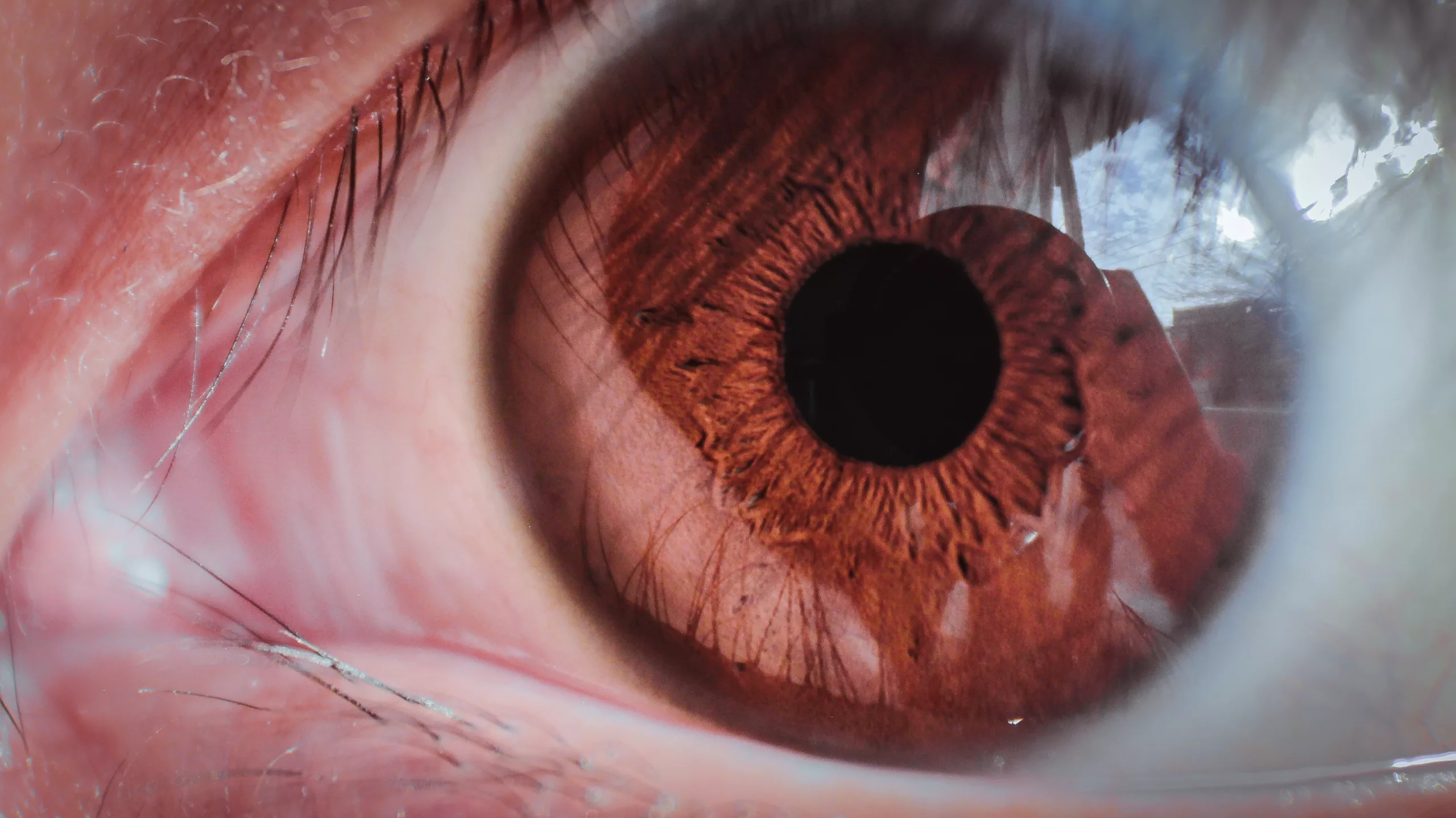
Gonioscopy is a diagnostic procedure that enables visualization of the iridocorneal angle using a gonioscope—a specialized lens system coupled with a light source. By placing the gonioscope directly onto the patient’s cornea, clinicians can examine the angle structures, including the trabecular meshwork, Schlemm’s canal, iris configuration, and peripheral anterior chamber.
Principles of Gonioscopy
The primary goal of gonioscopy is to assess the angle anatomy and identify any abnormalities or pathologies that may contribute to angle-closure or open-angle glaucoma. This examination technique relies on the principles of total internal reflection, which allows visualization of angle structures by directing light into the anterior chamber at various angles.
The fundamental principle underlying gonioscopy is total internal reflection of light. When light passes from a denser medium (such as the cornea) to a less dense medium (such as the aqueous humor), it undergoes reflection if the angle of incidence exceeds a critical angle. By directing a narrow beam of light onto the cornea at different angles, gonioscopy enables clinicians to visualize the angle structures and evaluate their morphology and integrity.
The Gonioscopic Procedure
Before performing gonioscopy, the patient’s eye is typically anesthetized with topical anesthetic drops to ensure comfort during the examination. A coupling agent, such as methylcellulose or goniosol, is then applied to the cornea to improve optical clarity and facilitate proper contact between the gonioscope lens and the eye. The clinician carefully places the gonioscope onto the cornea and systematically rotates it to examine all quadrants of the angle, ensuring a comprehensive assessment.
Types of Gonioscopy
- Direct Gonioscopy: Direct gonioscopy involves direct visualization of the angle structures using a handheld or slit-lamp-mounted gonioscope. This technique offers high-resolution images and allows for detailed examination of the angle morphology, pigmentation, and pathology. However, it requires skill and cooperation from the patient to maintain proper alignment and focus.
- Indirect Gonioscopy: Indirect gonioscopy utilizes a lens system, such as a Goldmann or Zeiss lens, in conjunction with a gonioscopic mirror. This approach provides a wider field of view and is particularly useful for patients with small or shallow anterior chambers. Although it offers a less magnified view compared to direct gonioscopy, it allows for panoramic visualization of the angle and facilitates examination in patients with limited cooperation or difficult anatomical configurations.
Applications in Ocular Health
Gonioscopy has numerous applications in the assessment and management of ocular conditions:
- Glaucoma Diagnosis: Gonioscopy is indispensable for differentiating between open-angle and angle-closure glaucoma. By evaluating the angle configuration, presence of peripheral anterior synechiae, and degree of trabecular meshwork pigmentation, clinicians can determine the underlying mechanism of glaucoma and tailor treatment strategies accordingly.
- Angle Assessment: Gonioscopy enables clinicians to assess the width and depth of the iridocorneal angle, detect signs of angle recession or neovascularization, and identify structural abnormalities such as iridotomy or iridectomy sites. This comprehensive evaluation aids in characterizing the angle anatomy and identifying potential risk factors for angle closure or glaucoma progression.
- Glaucoma Management: In the management of glaucoma, gonioscopy plays a critical role in monitoring disease progression, assessing the success of surgical interventions (such as trabeculectomy or laser trabeculoplasty), and guiding treatment adjustments to optimize intraocular pressure control and preserve visual function. Regular gonioscopic examinations allow for early detection of angle changes and proactive intervention to prevent vision loss.
Significance of Gonioscopy in Glaucoma Management
Gonioscopy is essential for optimizing glaucoma care and preserving vision in affected individuals. By providing clinicians with direct visualization of the iridocorneal angle, this technique facilitates accurate diagnosis, personalized treatment planning, and proactive management of glaucoma. By identifying anatomical abnormalities, evaluating angle dynamics, and monitoring changes over time, gonioscopy enables clinicians to tailor treatment strategies to address specific pathophysiological factors contributing to elevated intraocular pressure and glaucomatous damage.
Conclusion
In conclusion, gonioscopy serves as an indispensable tool in the diagnosis, management, and monitoring of glaucoma and other angle-related conditions. By offering clinicians direct access to the iridocorneal angle and facilitating comprehensive evaluation of angle structures, this technique plays a pivotal role in optimizing patient outcomes and preserving visual function. As our understanding of ocular anatomy and glaucoma pathophysiology continues to evolve, gonioscopy remains an essential component of the ophthalmologist’s armamentarium, ensuring timely intervention and personalized care for individuals at risk of glaucomatous damage.
World Eye Care Foundation’s eyecare.live brings you the latest information from various industry sources and experts in eye health and vision care. Please consult with your eye care provider for more general information and specific eye conditions. We do not provide any medical advice, suggestions or recommendations in any health conditions.
Commonly Asked Questions
Gonioscopy allows early detection of angle changes, facilitating proactive intervention to prevent vision loss and optimizing glaucoma care.
Gonioscopy is crucial for diagnosing and differentiating between different types of glaucoma by evaluating angle structures and pigmentations.
Yes, gonioscopy enables detection of angle abnormalities like recession or neovascularization, aiding in assessing glaucoma risk factors.
Gonioscopy helps monitor disease progression, assess surgical interventions’ success, and guide treatment adjustments for better glaucoma control.
Direct gonioscopy offers high-resolution images but requires skill and cooperation, while indirect gonioscopy provides a wider view and is useful for patients with difficult anatomies.
Before gonioscopy, the eye is typically anesthetized, and a coupling agent is applied to the cornea for better optical clarity.
Gonioscopy helps in tailoring treatment strategies by identifying anatomical abnormalities and monitoring changes over time to optimize intraocular pressure control.
Indirect gonioscopy provides a wider field of view, making it useful for patients with shallow anterior chambers or limited cooperation.
By assessing angle configuration, presence of synechiae, and trabecular meshwork pigmentation, gonioscopy aids in distinguishing between the two types of glaucoma.
Gonioscopy allows visualization of structures like the trabecular meshwork, Schlemm’s canal, and peripheral anterior chamber.


news via inbox
Subscribe here to get latest updates !


