
Inside Look: Mastering Pars Plana Vitrectomy
Introduction
Pars plana vitrectomy (PPV) is a sophisticated surgical procedure used to treat various eye conditions that affect the vitreous and retina. This guide offers an in-depth exploration of the procedure, covering its indications, techniques, potential complications, postoperative care, and the latest advancements in the field.
What is Pars Plana Vitrectomy?
Pars plana vitrectomy is a type of eye surgery that involves the removal of the vitreous gel from the eye. The vitreous is a clear, gel-like substance that fills the space between the lens and the retina. The microsurgical procedure is named after the pars plana, the flat part of the ciliary body, which is the preferred site for surgical entry to avoid damaging the lens and retina.
Indications for Pars Plana Vitrectomy
PPV is indicated for a variety of retinal and vitreous conditions, including:
- Retinal Detachment: A medical emergency where the retina peels away from its underlying layer of support tissue. If not treated promptly, it can lead to permanent vision loss.
- Vitreous Hemorrhage: Bleeding into the vitreous cavity, often caused by diabetic retinopathy or retinal vein occlusion. The blood can obscure vision and needs to be cleared to restore sight.
- Macular Hole: A small break in the macula, the central part of the retina responsible for detailed vision. It can cause central vision loss and distortion.
- Epiretinal Membrane: A thin layer of scar tissue that forms on the surface of the retina, causing vision to become blurred and distorted.
- Diabetic Retinopathy: Advanced stages of this condition can lead to severe retinal damage and vitreous hemorrhage due to abnormal blood vessel growth and leakage.
- Intraocular Infections: Severe infections such as endophthalmitis may require vitrectomy to remove infectious material and administer antibiotics directly into the eye.
- Retained Lens Fragments: Following cataract surgery, any remaining lens fragments in the vitreous cavity may need to be removed to prevent inflammation and further complications.
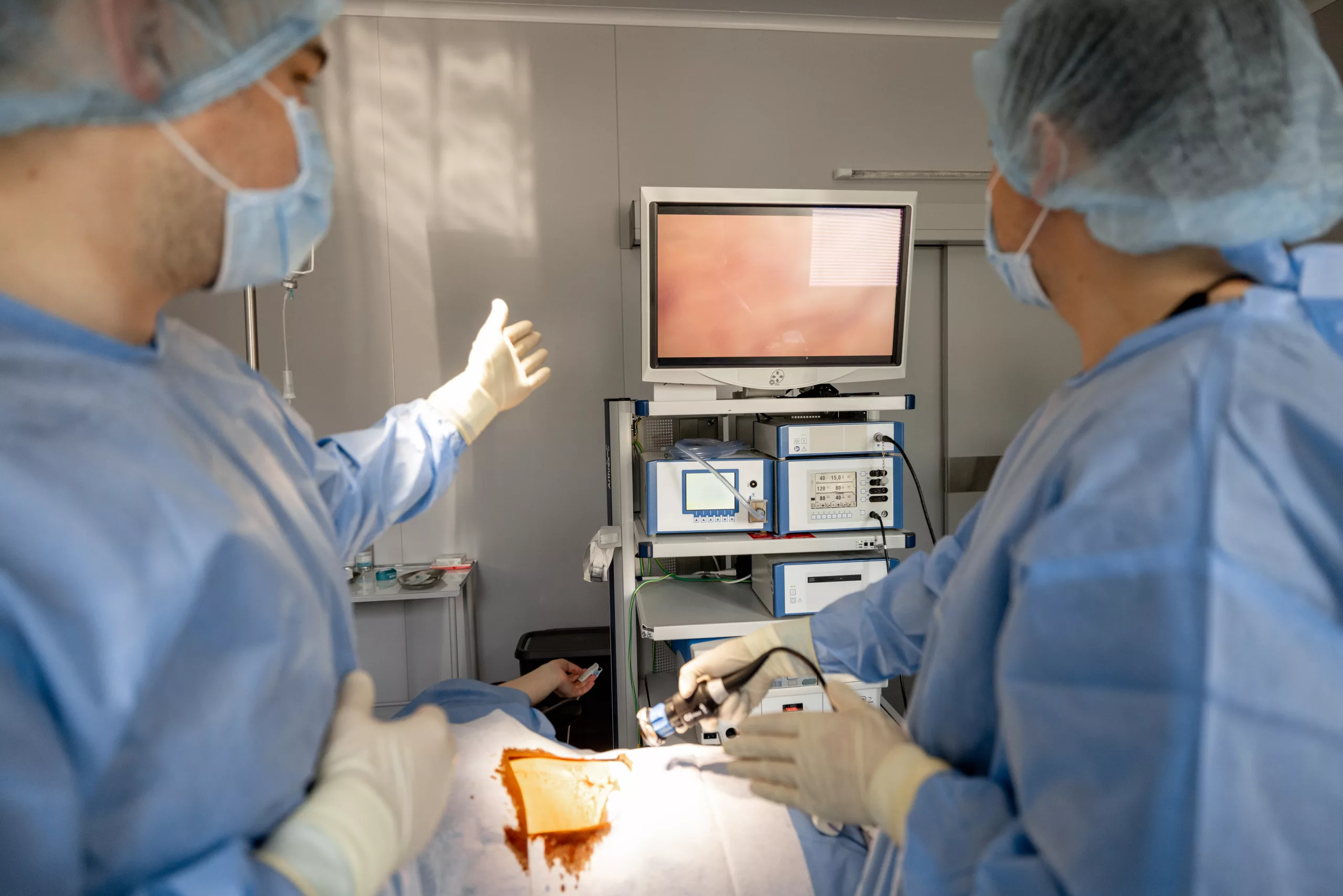
The Pars Plana Vitrectomy Procedure
PPV is typically performed under local or general anesthesia and involves several key steps:
- Preoperative Assessment: A thorough examination and imaging studies such as optical coherence tomography (OCT) and ultrasound are conducted to plan the surgery.
- Incisions: Small incisions (usually 1-2 millimeters) are made in the pars plana, located 3-4 millimeters behind the corneal limbus. This area provides safe access to the vitreous cavity without interfering with the lens or retina.
- Instrument Insertion: Specialized instruments, including a vitrector (a tiny cutting and suction device), an infusion cannula to maintain eye pressure, and a light source, are inserted through the incisions.
- Vitreous Removal: The vitreous gel is carefully removed to provide clear access to the retina. This step allows the surgeon to address the underlying condition, such as repairing retinal tears or removing scar tissue.
- Additional Procedures: Depending on the specific condition, the surgeon may perform additional procedures such as laser photocoagulation to seal retinal tears, peeling of epiretinal membranes, or injection of medication.
- Fluid-Gas Exchange: In some cases, a gas bubble or silicone oil is injected into the eye to help flatten the retina against the back of the eye and promote healing. The type of gas or oil used depends on the condition being treated and the surgeon’s preference.
Postoperative Care and Recovery
After PPV, patients need to follow specific postoperative care instructions to ensure proper healing and avoid complications:
- Positioning: If a gas bubble is used, patients may need to maintain a specific head position (such as face-down) for a certain period to keep the bubble in contact with the retina. This positioning helps support the retina as it heals.
- Activity Restrictions: Strenuous activities and heavy lifting should be avoided during the initial recovery period to prevent increased eye pressure and other complications.
- Medications: Patients are usually prescribed a regimen of eye drops, including antibiotics to prevent infection, steroids to reduce inflammation, and sometimes medications to control intraocular pressure.
- Follow-Up Visits: Regular follow-up appointments with the ophthalmologist are essential to monitor the healing process, check for any signs of complications, and ensure that the retina remains attached.
Potential Complications
While PPV is generally safe and effective, it carries some risks and potential complications:
- Infection: Though rare, infections can occur and may require additional treatment. Sterile techniques and postoperative antibiotics are used to minimize this risk.
- Bleeding: Intraocular bleeding during or after surgery can affect vision and may require further intervention.
- Cataract Formation: The procedure can accelerate the development of cataracts, particularly in older patients, necessitating cataract surgery at a later stage.
- Retinal Detachment: Despite the intent to repair retinal detachment, new detachments can occur postoperatively, requiring further surgical intervention.
- Increased Intraocular Pressure: Elevated eye pressure can develop postoperatively and may need to be managed with medications or additional surgery.
- Recurrent Macular Hole: In some cases, macular holes may reopen and require another surgical procedure.
Advances in Vitrectomy Techniques
Technological advancements have significantly improved vitrectomy techniques and outcomes:
- Small-Gauge Surgery: The use of smaller instruments (23-, 25-, and 27-gauge) allows for less invasive surgery, faster recovery, and fewer complications. Smaller incisions often do not require sutures, reducing postoperative discomfort and inflammation.
- High-Speed Vitrectors: Modern vitrectors operate at higher speeds, allowing for safer and more efficient vitreous removal. These devices can cut the vitreous gel into smaller pieces, minimizing traction on the retina.
- Enhanced Visualization: Advanced imaging techniques, such as optical coherence tomography (OCT) and intraoperative optical coherence tomography (iOCT), provide real-time, high-resolution images of the retina during surgery. These tools help surgeons make precise decisions and improve surgical outcomes.
- Improved Illumination Systems: Modern illumination systems provide better visualization of the retina and vitreous, enhancing the surgeon’s ability to perform delicate procedures accurately.
Conclusion
Pars plana vitrectomy is a vital surgical procedure for treating various serious retinal and vitreous conditions. With advances in surgical techniques and technologies, PPV has become safer and more effective, offering improved outcomes for patients. If you are experiencing symptoms of retinal disease or have been advised to undergo vitrectomy, consulting with a retinal specialist can provide you with the necessary information and guidance for your condition.
By understanding the procedure, its indications, potential complications, and advancements, patients can make informed decisions about their eye health and treatment options.
World Eye Care Foundation’s eyecare.live brings you the latest information from various industry sources and experts in eye health and vision care. Please consult with your eye care provider for more general information and specific eye conditions. We do not provide any medical advice, suggestions or recommendations in any health conditions.
Commonly Asked Questions
PPV is recommended for conditions like retinal detachment, vitreous hemorrhage (due to diabetic retinopathy or trauma), macular hole, epiretinal membrane, and complications following cataract surgery such as retained lens fragments.
No, PPV is performed under anesthesia, either local or general, ensuring that patients do not experience pain during the surgery. Mild discomfort or pressure may be felt, but pain is typically minimal.
Recovery time varies, but most patients can resume normal activities within a few days to weeks after surgery. Complete recovery, including stabilization of vision, may take several weeks to months, depending on the individual case.
Risks include infection, bleeding inside the eye, increased intraocular pressure, cataract formation, and, in rare cases, retinal detachment. These risks are mitigated through careful preoperative evaluation and postoperative care.
While uncommon, complications such as infection, persistent bleeding, or recurrence of retinal conditions like macular hole or epiretinal membrane can occur. Regular follow-up visits with an ophthalmologist are crucial to monitor for any signs of complications.
Most PPV procedures are performed on an outpatient basis, allowing patients to return home the same day. Rarely, overnight hospitalization may be required for monitoring or if there are specific medical considerations.
Patients should not drive until their vision has stabilized and they feel comfortable with their visual acuity. This may vary from a few days to several weeks depending on individual healing and the advice of the treating ophthalmologist.
The need for glasses after PPV depends on factors such as the patient’s pre-existing vision, the specific condition treated, and the use of intraocular tamponades (gas or oil). Some patients may require new prescriptions due to changes in their eye’s refractive status post-surgery.
Initially, patients are advised to avoid strenuous activities, heavy lifting, or activities that increase intraocular pressure. Gradually, normal activities can be resumed as directed by the ophthalmologist during follow-up visits.
Yes, in some cases, repeat PPV may be necessary if complications recur or new conditions develop. The decision for repeat surgery is made based on the individual patient’s response to initial treatment and ongoing retinal health.


news via inbox
Subscribe here to get latest updates !


