
Ophthalmological Evaluation by a Maxillofacial Surgeon
Introduction
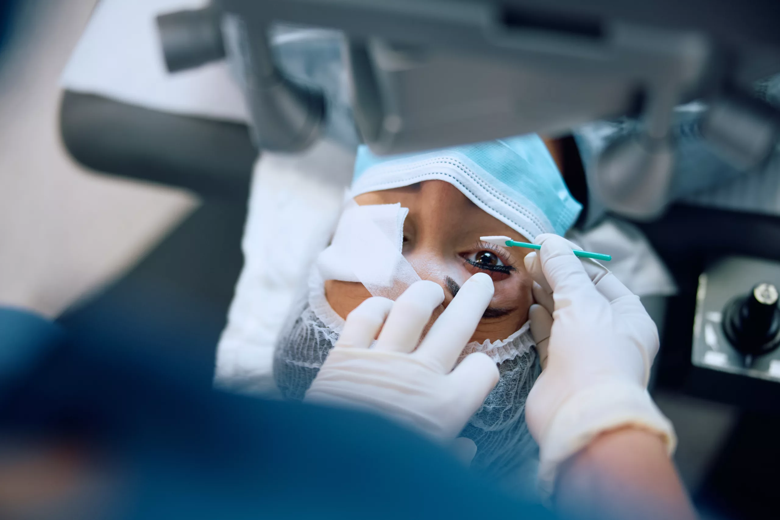
An ophthalmological evaluation by a maxillofacial surgeon is pivotal in addressing complex conditions affecting the eyes and facial structures. This specialized evaluation combines expertise in craniofacial anatomy with surgical proficiency, ensuring comprehensive care for patients with traumatic injuries, congenital anomalies, tumors, and other pathologies impacting the ocular region.
Scope of Practice for Maxillofacial Surgeons in Ophthalmology
Maxillofacial surgeons possess a broad scope in ophthalmology, encompassing:
- Craniofacial Trauma: Maxillofacial surgeons are adept at managing traumatic injuries involving the orbits (eye sockets), eyelids, and adjacent facial bones. This includes fractures resulting from accidents or assaults, where precise reconstruction is crucial for preserving both functional vision and facial aesthetics.
- Congenital Anomalies: They diagnose and treat congenital conditions affecting the ocular region, such as craniosynostosis (premature fusion of skull bones) or cleft lip and palate. Early intervention is critical to address associated ocular complications like strabismus (misalignment of the eyes) or refractive errors.
- Tumors and Pathologies: Maxillofacial surgeons play a key role in the management of tumors affecting the eye, orbit, or adjacent soft tissues. This may involve surgical excision, biopsy, or reconstruction to achieve optimal outcomes while preserving vision and facial symmetry.
- Reconstructive Surgery: Following trauma or surgical interventions, maxillofacial surgeons restore facial function and aesthetics. This includes eyelid reconstruction to correct ptosis (drooping eyelid) or ectropion (outward turning of the eyelid), ensuring normal eyelid function and protection of the ocular surface.
Key Components of Ophthalmological Evaluation
- Patient History and Physical Examination:
- Comprehensive History: Involves gathering detailed information about ocular symptoms, previous surgeries, medical history (including systemic diseases impacting ocular health), and family history of eye disorders. This information guides diagnostic decisions and treatment planning.
- Physical Examination: A thorough assessment includes:
- Visual Acuity: Measurement of central vision using standard eye charts (Snellen chart) or more specialized tests (e.g., ETDRS charts for research or clinical trials).
- Pupillary Reactions: Evaluation of pupil size, shape, and response to light, assessing both direct and consensual responses for neurological integrity.
- Extraocular Movements: Examination of eye movements in all cardinal directions of gaze, identifying limitations or abnormalities that may indicate cranial nerve dysfunction.
- Facial Symmetry and Sensation: Assessment of facial nerve function (cranial nerve VII), evaluating symmetry of facial expressions and sensation (e.g., touch, pain) in the periorbital region.
- Diagnostic Imaging and Tests:
- CT Scan and MRI: Imaging modalities provide detailed anatomical visualization of facial and orbital structures. CT scans are particularly useful for assessing bony integrity and fractures, while MRI offers superior soft tissue contrast, aiding in the diagnosis of orbital tumors or vascular malformations.
- Ocular Ultrasonography: This non-invasive technique uses high-frequency sound waves to visualize intraocular structures (e.g., retina, optic nerve) and assess abnormalities such as retinal detachment or intraocular tumors. It is particularly valuable when direct visualization with conventional ophthalmic instruments is limited.
- Specialized Assessments:
- Visual Field Testing: Evaluates the full horizontal and vertical range of peripheral vision, detecting abnormalities indicative of glaucoma, optic nerve diseases, or neurological disorders affecting visual pathways.
- Optical Coherence Tomography (OCT): A non-invasive imaging technique that provides cross-sectional images of the retina and optic nerve head. It is instrumental in diagnosing and monitoring conditions such as macular degeneration, diabetic retinopathy, and glaucoma by measuring retinal thickness and detecting subtle structural changes over time.
- Collaboration with Ophthalmologists:
- Multidisciplinary Approach: Maxillofacial surgeons collaborate closely with ophthalmologists to integrate surgical and medical management plans. This ensures comprehensive care, especially in complex cases requiring combined expertise in craniofacial surgery and ophthalmology.
- Postoperative Care: Following surgical interventions affecting the ocular region, maxillofacial surgeons coordinate postoperative care to optimize recovery and functional outcomes. This may involve monitoring for complications such as infection, hematoma, or delayed wound healing, and ensuring patient education regarding postoperative care instructions and potential visual outcomes.
Common Conditions Evaluated and Treated
- Orbital Fractures: These fractures may involve the orbital floor, walls, or roof, potentially leading to ocular mobility restrictions, diplopia (double vision), or enophthalmos (sunken eye). Maxillofacial surgeons employ precise surgical techniques (e.g., orbital reconstruction with implants or bone grafts) to restore orbital anatomy and function while minimizing long-term sequelae.
- Eyelid Disorders: Conditions such as ptosis (drooping eyelid), entropion (inward turning of the eyelid), or ectropion (outward turning of the eyelid) can affect eyelid function, ocular surface health, and visual acuity. Surgical correction by maxillofacial surgeons restores eyelid position and function, improving both aesthetics and ocular protection.
- Orbital Tumors: Benign or malignant tumors affecting the orbit require accurate diagnosis and surgical intervention to achieve complete excision while preserving vision and orbital integrity. Maxillofacial surgeons work in conjunction with oncologists and ophthalmologists to develop individualized treatment plans, incorporating surgical resection, radiation therapy, or chemotherapy as indicated.
Advances in Ophthalmological Evaluation by Maxillofacial Surgeons
- Technology Integration: The integration of advanced imaging modalities (e.g., 3D CT scanning, intraoperative navigation systems) enhances diagnostic accuracy and surgical precision. This allows for minimally invasive approaches and customized treatment strategies tailored to each patient’s unique anatomy and pathology.
- Patient-Centered Care: Maxillofacial surgeons prioritize patient-centered care, emphasizing informed consent, shared decision-making, and ongoing communication throughout the treatment continuum. This holistic approach ensures that patient preferences and expectations are addressed, promoting overall satisfaction and optimizing long-term outcomes.
Conclusion
An ophthalmological evaluation by a maxillofacial surgeon represents a cornerstone in the management of complex craniofacial and ocular conditions. By leveraging their expertise in facial anatomy, surgical proficiency, and collaborative approach with ophthalmologists, these specialists ensure comprehensive care from diagnosis through rehabilitation. Through ongoing advancements in technology and patient-centered care principles, maxillofacial surgeons continue to enhance outcomes and quality of life for patients requiring specialized ocular evaluation and treatment.
World Eye Care Foundation’s eyecare.live brings you the latest information from various industry sources and experts in eye health and vision care. Please consult with your eye care provider for more general information and specific eye conditions. We do not provide any medical advice, suggestions or recommendations in any health conditions.
Commonly Asked Questions
Patients should gather relevant medical records, including previous surgeries, medications, and any known allergies. They should also be prepared to discuss symptoms, concerns, and goals for treatment during the initial consultation. Following preoperative instructions and maintaining good overall health can support successful outcomes.
Yes, maxillofacial surgeons with expertise in pediatric craniofacial surgery collaborate with pediatric ophthalmologists to evaluate and treat congenital eye conditions in children. Early intervention is crucial to address developmental abnormalities and optimize visual outcomes.
Recovery time varies depending on the type of surgery performed and individual patient factors. Patients may experience swelling, bruising, and discomfort initially, which typically subsides within a few weeks. Maxillofacial surgeons provide specific postoperative instructions to promote healing and optimize recovery.
Risks and complications may include infection, bleeding, damage to nearby structures (e.g., nerves or blood vessels), and aesthetic concerns. However, careful preoperative planning, precise surgical techniques, and comprehensive postoperative care minimize these risks and optimize outcomes.
Depending on the type and location of the tumor, maxillofacial surgeons may collaborate with oncologists and ophthalmologists to consider non-surgical treatments such as radiation therapy or chemotherapy. These treatments aim to shrink or control tumor growth while preserving vision and orbital function.
Maxillofacial surgeons utilize advanced imaging modalities such as CT scans and MRI to visualize facial and orbital structures in detail. These imaging techniques help in diagnosing fractures, tumors, vascular malformations, and other pathologies affecting the eye and surrounding tissues.
Yes, maxillofacial surgeons are trained to perform eyelid surgery (blepharoplasty) to correct functional or cosmetic issues affecting the eyelids. This may include correcting ptosis (drooping eyelid), entropion (inward turning of the eyelid), or addressing eyelid deformities following trauma or congenital anomalies.
Conditions such as orbital fractures, tumors affecting the eye or orbit, congenital anomalies like craniosynostosis, and complex facial trauma involving the ocular region may necessitate specialized evaluation and treatment by a maxillofacial surgeon with expertise in ophthalmology.
Maxillofacial surgeons and ophthalmologists work closely to develop comprehensive treatment plans for patients. This collaboration involves sharing diagnostic findings, discussing surgical options, and coordinating postoperative care to ensure optimal outcomes for conditions affecting both the facial and ocular structures.
Maxillofacial surgeons undergo extensive training in both medicine and dentistry, followed by specialized residency training focusing on surgical procedures of the face, jaws, and associated structures. They often collaborate with ophthalmologists to manage complex conditions affecting the eyes and orbital region.


news via inbox
Subscribe here to get latest updates !


