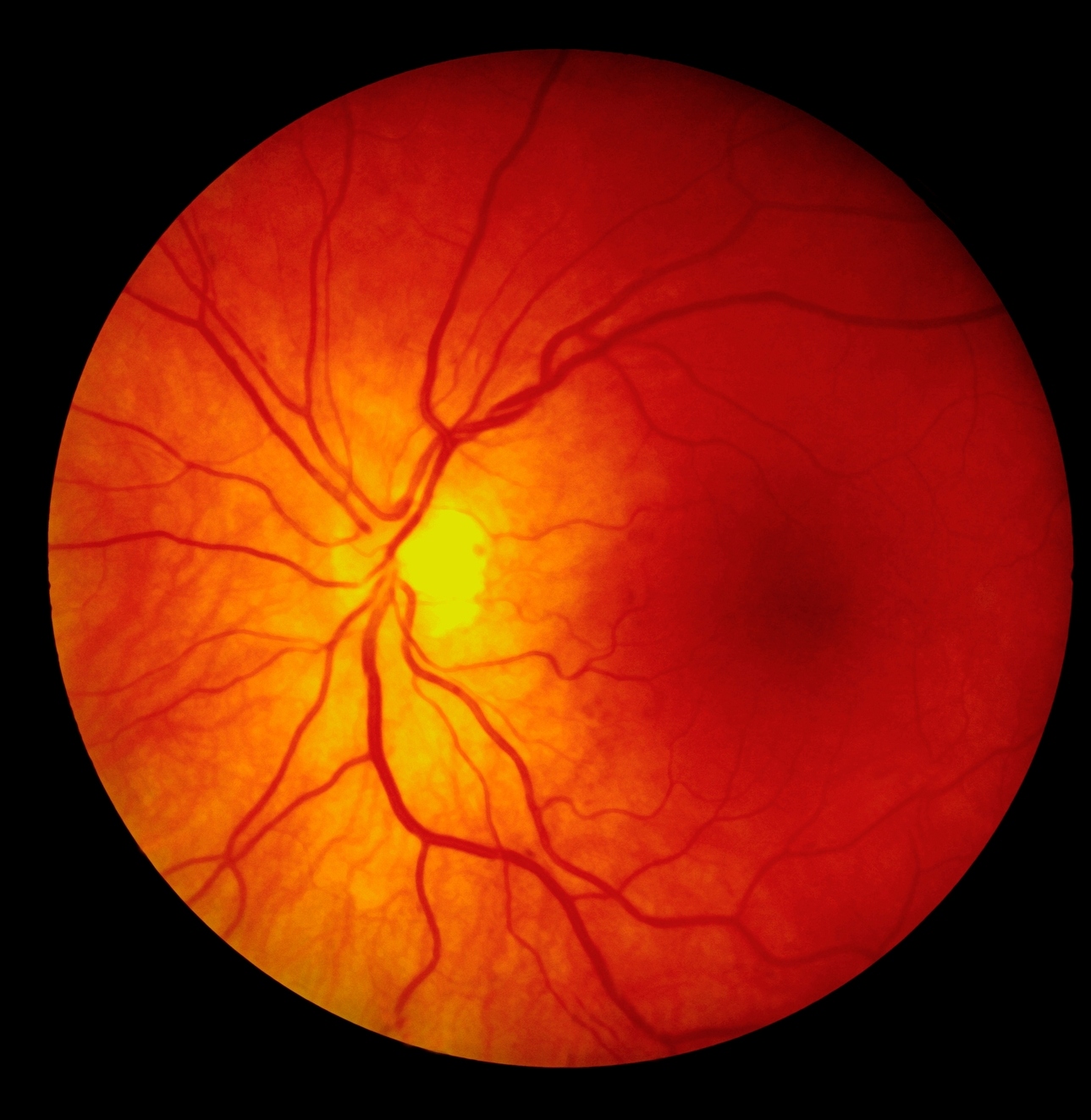
Macular Hole: Diagnosis and Surgical Intervention
Understanding Macular Holes
Macular holes are a type of eye condition that can affect vision, particularly central vision. To understand macular holes, it’s important to know about the macula, which is a small area in the retina at the back of the eye responsible for central vision and detailed sight.
When a macular hole develops, it means there is a small break or opening in the macula. This can lead to distorted or blurry vision, making it difficult to see fine details or to focus on objects directly in front of you.
Symptoms of macular holes typically include
- Blurred or Distorted Vision: One of the most common symptoms of a macular hole is blurry or distorted central vision. This can make it challenging to read, drive, or recognize faces.
- Straight Lines Appearing Wavy: Another symptom is that straight lines may appear distorted or wavy when looking at them directly.
- Dark or Empty Area in Vision: Some people with macular holes may experience a dark or empty area in the center of their vision. This can create a sensation of a blind spot or a hole in the vision.
- Difficulty with Low Light Vision: Macular holes can also make it harder to see in low light conditions, such as at night or in dimly lit rooms.
- Decreased Visual Acuity: As the macular hole progresses, there may be a noticeable decrease in visual acuity, affecting activities that require clear central vision.
Causes
Macular holes typically develop as a result of changes that occur in the vitreous, a gel-like substance that fills the inside of the eye. As we age, the vitreous can shrink and pull away from the surface of the retina. This process, known as vitreous detachment, can sometimes exert traction on the macula, leading to the formation of a hole.
Other factors that may contribute to the development of macular holes include:
- Age: Macular holes are more common in people over the age of 60.
- Eye Trauma: Injury to the eye can increase the risk of developing a macular hole.
- Eye Conditions: Certain eye conditions, such as high myopia (severe nearsightedness) or retinal detachment, can predispose individuals to macular holes.
- Eye Surgery: Previous eye surgeries, such as cataract surgery or vitrectomy, may also increase the risk of macular holes.
Risk Factors
Several factors can increase a person’s risk of developing macular holes, including:
- Age: As mentioned, macular holes are more prevalent in older adults, particularly those over the age of 60.
- Eye Conditions: People with existing eye conditions, such as diabetic retinopathy or macular pucker, may be at a higher risk.
- High Myopia: Severe nearsightedness is associated with an increased risk of macular holes.
- Eye Trauma: Any injury to the eye can potentially lead to the development of macular holes.
Diagnosis of Macular Holes
Diagnosing a macular hole typically involves a comprehensive eye examination by an eye care professional. The following steps are usually involved:
- Visual Acuity Test: This involves reading letters on an eye chart to assess central vision.
- Dilated Eye Exam: The eye doctor will use special eye drops to dilate the pupils, allowing for a detailed examination of the retina and macula.
- Optical Coherence Tomography (OCT): This imaging test provides high-resolution cross-sectional images of the retina, allowing the doctor to visualize any abnormalities, including macular holes.
- Fundus Photography: Photographs of the back of the eye may be taken to document the appearance of the macular hole and monitor changes over time.
- Amsler Grid Test: This test involves looking at a grid of straight lines to detect any distortions or gaps in vision that may indicate a macular hole.
Surgical Intervention Options
- Vitrectomy: A vitrectomy is a surgical procedure where the vitreous gel inside the eye is removed and replaced with a clear solution. During the procedure, the surgeon may also perform additional steps such as removing the internal limiting membrane (ILM) and injecting a gas bubble into the eye to help close the macular hole.
- ILM Peeling: Removing the ILM, a thin membrane on the surface of the retina, can help release traction on the macula and improve the success rate of macular hole closure.
- Gas Tamponade: After surgery, a gas bubble may be injected into the eye to provide internal support and facilitate the closure of the macular hole. The patient may need to maintain a face-down position for a period to ensure proper positioning of the gas bubble.
Recovery and Rehabilitation
- Recovery Process: The recovery process following macular hole surgery typically involves gradual improvement in vision over several weeks to months. Patients may need to refrain from strenuous activities and follow postoperative instructions provided by their surgeon.
- Expected Outcomes and Complications: While most patients experience improved vision following surgery, complications such as cataract formation, elevated eye pressure, or recurrent macular holes may occur in some cases.
- Rehabilitation Strategies: Visual aids and rehabilitation exercises may be recommended to help patients adapt to any remaining visual deficits and optimize their vision post-surgery.
Advances in Surgical Techniques and Research
- Innovations: Ongoing research and technological advancements continue to refine surgical techniques and improve outcomes for patients with macular holes. This may include advancements in imaging technology, surgical instrumentation, and intraocular implants.
- Future Directions: Emerging treatment modalities such as pharmacologic agents or gene therapy hold promise for further enhancing the management of macular holes and related retinal conditions.
Conclusion
In conclusion, understanding the diagnosis and surgical intervention options for macular holes is crucial for both healthcare professionals and patients alike. Through advancements in technology and surgical techniques, the prognosis for individuals with macular holes has significantly improved. However, early detection and prompt treatment remain key to preserving vision and optimizing outcomes. By staying informed about the latest developments in the field, individuals can make informed decisions about their eye health and seek appropriate care when needed.
World Eye Care Foundation’s eyecare.live brings you the latest information from various industry sources and experts in eye health and vision care. Please consult with your eye care provider for more general information and specific eye conditions. We do not provide any medical advice, suggestions or recommendations in any health conditions.
Commonly Asked Questions
Following macular hole surgery, patients typically have scheduled follow-up appointments with their eye surgeon to monitor the progress of healing and assess visual acuity. These appointments may involve additional imaging tests, such as OCT, to evaluate the status of the macular hole closure.
While macular holes cannot always be prevented, maintaining good eye health and attending regular eye exams can help detect any early signs of macular holes or other retinal conditions. Prompt treatment of conditions such as diabetic retinopathy or retinal tears may also reduce the risk of developing macular holes.
While surgery is the primary treatment for macular holes, certain pharmacologic agents, such as ocriplasmin, have been explored as potential non-surgical alternatives. However, these treatments are not as effective as surgery and may be reserved for select cases.
Complications of macular hole surgery may include cataract formation, elevated intraocular pressure, retinal detachment, or persistent macular hole despite surgical intervention. However, these complications are relatively rare and can often be managed with appropriate follow-up care.
While macular hole surgery can significantly improve vision for many patients, the extent of visual recovery depends on factors such as the size and duration of the macular hole, as well as any preexisting retinal damage. Some individuals may experience a partial restoration of vision, while others may achieve near-normal visual acuity.
While macular hole surgery is successful in closing the hole in the majority of cases, there is a small risk of recurrence. Factors such as the size and stage of the macular hole, as well as the surgical technique used, can influence the likelihood of recurrence.
While there are no specific lifestyle changes required after macular hole surgery, patients are often advised to avoid activities that may put undue strain on the eyes, such as heavy lifting or strenuous exercise, during the initial recovery period.
The recovery period following macular hole surgery can vary from several weeks to months. During this time, vision gradually improves as the eye heals, and patients may need to follow specific postoperative instructions provided by their surgeon.
Macular hole surgery is typically performed under local anesthesia, so patients may experience some discomfort or pressure during the procedure. However, postoperative pain is usually minimal and can be managed with medications prescribed by your surgeon.
While some smaller macular holes may spontaneously close without surgical intervention, larger or persistent macular holes typically require surgical treatment to prevent vision loss.
news via inbox
Subscribe here to get latest updates !


