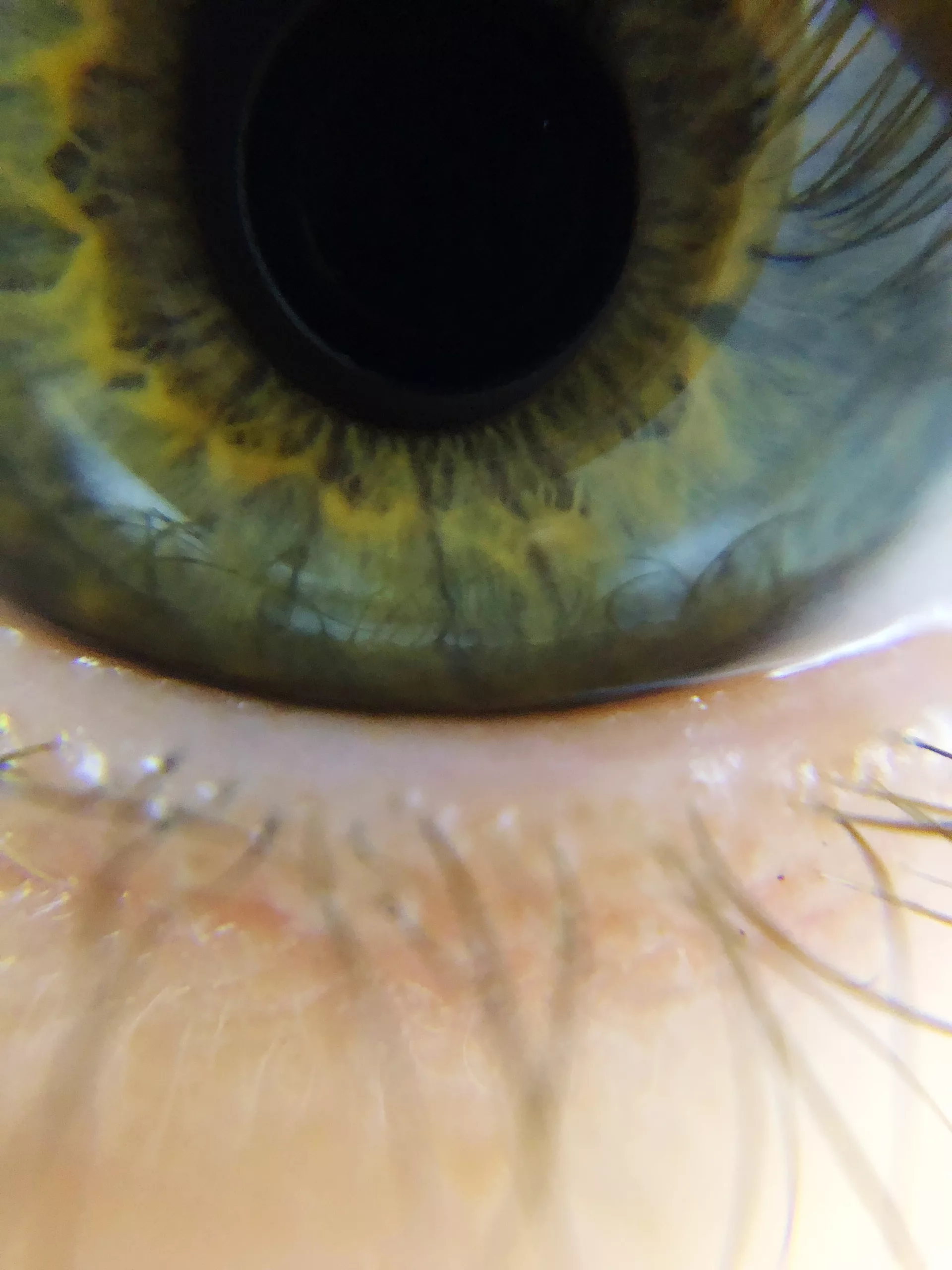
Palpebral Fissure Asymmetry: In-Depth Analysis
Introduction
Palpebral fissure asymmetry refers to the condition where there is a noticeable difference in the size, shape, or position of the openings between the eyelids of each eye. This condition, while often a minor cosmetic concern, can sometimes signal underlying medical issues, impact vision, and affect the overall symmetry of the face. Below is a more detailed exploration of this topic, covering every aspect of palpebral fissure asymmetry.
Anatomy of the Palpebral Fissure
The palpebral fissure is the elliptical opening between the upper and lower eyelids. It typically measures about 10-12 mm vertically and 25-30 mm horizontally in adults. The symmetry of the palpebral fissure is largely determined by the position and function of the eyelids, which are controlled by the orbicularis oculi muscle, the levator palpebrae superioris muscle, and the tarsal plates.
Anatomical Components Influencing the Palpebral Fissure
- Eyelids: The upper and lower eyelids, composed of skin, muscle, and connective tissue, frame the palpebral fissure. The eyelids are responsible for protecting the eyes, maintaining moisture, and controlling the amount of light that enters the eyes.
- Levator Palpebrae Superioris Muscle: This muscle is primarily responsible for lifting the upper eyelid. Proper functioning of this muscle is essential for maintaining the normal position of the upper eyelid, contributing to the symmetry of the palpebral fissure.
- Orbicularis Oculi Muscle: This circular muscle encircles the eye and is responsible for closing the eyelids. It works in coordination with the levator muscle to maintain the appropriate position and function of the eyelids.
- Tarsal Plates: These are dense connective tissue structures within the eyelids that provide structural support and shape. The tarsal plates help maintain the contour of the eyelids and, consequently, the shape of the palpebral fissure.
Importance of Palpebral Fissure Symmetry
Symmetry in the palpebral fissure is important not only for aesthetics but also for ocular health. Even minor asymmetries can alter the distribution of tear film across the eye, potentially leading to dry eyes or other ocular surface disorders. Additionally, significant asymmetry can impact vision by obstructing the visual axis.
Causes of Palpebral Fissure Asymmetry
Palpebral fissure asymmetry can result from a wide range of conditions, both congenital and acquired. Understanding these causes is critical for accurate diagnosis and effective treatment.
Congenital Ptosis
- Definition: Congenital ptosis is a condition present at birth where the upper eyelid droops due to underdevelopment or dysfunction of the levator palpebrae superioris muscle.
- Pathophysiology: The levator muscle may be structurally abnormal, leading to weak or incomplete elevation of the eyelid. This condition can be unilateral (affecting one eye) or bilateral (affecting both eyes) and often requires early surgical intervention to prevent amblyopia (lazy eye) or other vision problems.
Facial Nerve Palsy
- Definition: Facial nerve palsy is a condition in which the facial nerve (cranial nerve VII) is damaged, leading to weakness or paralysis of the muscles of facial expression, including those controlling the eyelids.
- Causes: Common causes include Bell’s palsy, trauma, tumors, or stroke. The resultant muscle weakness can cause the lower eyelid to droop or the upper eyelid to sag, leading to palpebral fissure asymmetry.
- Symptoms: Besides eyelid asymmetry, patients may experience difficulty blinking, drooling, or a lack of facial expression on the affected side.
Thyroid Eye Disease (Graves’ Ophthalmopathy)
- Definition: Thyroid eye disease (TED) is an autoimmune condition associated with hyperthyroidism, particularly Graves’ disease, where the immune system attacks the tissues around the eyes.
- Pathophysiology: In TED, the muscles that control the eyelids and the tissues around the eyes become inflamed and swollen, often leading to retraction of the upper eyelids. This retraction causes a wider and more open palpebral fissure, leading to noticeable asymmetry.
- Clinical Features: Patients may also present with proptosis (bulging eyes), double vision, and discomfort or pain around the eyes.
Trauma
- Description: Trauma to the face or eyes can lead to injuries such as fractures, lacerations, or burns that can disrupt the normal anatomy and function of the eyelids, resulting in asymmetry.
- Mechanisms: Trauma can cause scarring, damage to the levator muscle, or nerve injury, all of which can alter the position of the eyelids. The extent of the asymmetry depends on the severity and location of the trauma.
Blepharoptosis (Acquired Ptosis)
- Definition: Acquired ptosis refers to the drooping of the upper eyelid that occurs later in life due to various causes, including aging, muscle disorders, or nerve damage.
- Causes: The most common cause is age-related weakening of the levator aponeurosis (the tendon-like structure that connects the levator muscle to the eyelid). Other causes include neuromuscular diseases like myasthenia gravis, third nerve palsy, or mechanical issues such as tumors.
- Clinical Impact: Acquired ptosis can affect one or both eyes and may lead to visual field obstruction if the eyelid droops significantly over the pupil.
Dermatochalasis
- Definition: Dermatochalasis is the excess skin on the upper or lower eyelids that typically occurs due to aging. It can cause one eyelid to appear lower or more wrinkled than the other, leading to asymmetry.
- Symptoms: This condition often leads to a tired or heavy appearance and can contribute to visual impairment if the excess skin obstructs the visual axis.
Orbital Masses or Tumors
- Description: Growths or tumors within the orbit (the bony cavity that contains the eye) can exert pressure on the eyelids or the muscles controlling them, leading to asymmetry.
- Types of Masses: These can include benign conditions like cysts or hemangiomas, as well as malignant tumors such as lymphoma or metastatic disease. The mass effect can push the eyelid or eye itself out of position, creating noticeable asymmetry.
- Diagnosis and Treatment: Imaging studies are essential for diagnosis, and treatment typically involves surgical removal or other interventions to address the underlying mass.
Diagnosis of Palpebral Fissure Asymmetry
Accurate diagnosis of palpebral fissure asymmetry is critical for determining the underlying cause and planning appropriate treatment.
Visual Inspection
- Procedure: A thorough visual examination of the face, eyelids, and eyes is conducted to assess the symmetry of the palpebral fissures. The examiner observes the patient in various positions of gaze to evaluate the function of the eyelid muscles.
- Important Observations: Asymmetry in the vertical height of the fissures, differences in the position of the upper or lower eyelids, and any associated eyelid abnormalities (e.g., ptosis, retraction) are noted.
Measurement of the Palpebral Fissure
- Tools: Measurements are typically taken using a ruler or calipers to quantify the vertical and horizontal dimensions of the palpebral fissures on both eyes.
- Interpretation: Significant differences between the two eyes can indicate the presence of underlying conditions. For example, a marked difference in vertical height may suggest ptosis, while an increased horizontal width could indicate eyelid retraction.
Assessment of Eyelid Function
- Tests: The function of the levator palpebrae superioris and orbicularis oculi muscles is assessed through tests like the margin reflex distance (MRD), levator function test, and eyelid excursion measurements.
- Findings: Reduced levator function or decreased eyelid excursion can indicate ptosis, while abnormal orbicularis function may point to facial nerve palsy.
Assessment of Ocular Health
- Comprehensive Eye Exam: A complete eye examination is performed to rule out other ocular conditions that could be contributing to the asymmetry. This includes checking visual acuity, ocular surface health, and intraocular pressure.
- Imaging Studies: In cases where a structural abnormality is suspected, imaging studies such as MRI, CT scans, or ultrasound may be employed to visualize the orbit and surrounding tissues. These studies can help identify masses, structural deformities, or muscle abnormalities that could be causing the asymmetry.
Consideration of Systemic Health
- Systemic Evaluation: In cases where systemic diseases (e.g., thyroid eye disease, myasthenia gravis) are suspected, additional systemic evaluations and tests such as blood work or electrophysiological studies may be necessary.
Treatment Options for Palpebral Fissure Asymmetry
Treatment options for palpebral fissure asymmetry are tailored to the underlying cause and the severity of the asymmetry.
Surgical Correction
- Indications: Surgery is often indicated for significant asymmetry that affects vision, causes discomfort, or is cosmetically concerning. Conditions such as ptosis, dermatochalasis, and orbital tumors are commonly treated surgically.
- Common Procedures:
- Ptosis Surgery (Blepharoptosis Repair): This involves tightening or shortening the levator muscle or its aponeurosis to elevate the eyelid to a more symmetrical position.
- Blepharoplasty: This procedure involves the removal of excess skin, muscle, or fat from the eyelids, often performed for dermatochalasis.
- Orbitotomy: Surgical removal of orbital masses or tumors may be necessary to relieve pressure
and correct eyelid position.
Botulinum Toxin Injections
- Use: Botulinum toxin (e.g., Botox) is used to temporarily paralyze muscles. It can be beneficial in treating facial nerve palsy, thyroid eye disease, or other conditions where muscle overactivity or underactivity is contributing to asymmetry.
- Mechanism: Injections are administered to specific muscles to improve eyelid position and function. The effects are temporary, and repeated treatments may be necessary.
Management of Underlying Conditions
- Thyroid Eye Disease: Management includes controlling thyroid levels, using medications or corticosteroids to reduce inflammation, and in severe cases, surgical intervention to correct eyelid retraction.
- Myasthenia Gravis: Treatment involves anticholinesterase medications, immunosuppressants, and possibly thymectomy to improve muscle strength and reduce ptosis.
- Facial Nerve Palsy: Management may include physical therapy, medications, or surgical options depending on the severity and underlying cause of the nerve damage.
Prosthetics and Aids
- Eyelid Weights: Small weights can be attached to the upper eyelids to help keep them in a more open position, particularly useful in cases of ptosis or facial nerve weakness.
- Custom Prosthetics: In some cases, custom-made prosthetic devices can help achieve a more symmetrical appearance.
Prognosis and Outcomes
The prognosis for palpebral fissure asymmetry depends on the underlying cause and the effectiveness of the treatment.
Post-Treatment Outcomes
- Surgical Outcomes: Most patients experience significant improvement in eyelid position and symmetry following surgery. However, results can vary based on the individual’s specific condition and the complexity of the surgery.
- Long-Term Management: Some conditions, like thyroid eye disease or facial nerve palsy, may require ongoing management to maintain results and address any recurrence or progression.
- Quality of Life: Improvement in palpebral fissure symmetry can enhance both visual function and aesthetic appearance, contributing to better overall quality of life.
Additional Considerations
Psychosocial Impact
- Emotional and Social Effects: Significant palpebral fissure asymmetry can impact self-esteem and social interactions. Patients may experience psychological distress or anxiety related to their appearance.
- Support and Counseling: Providing emotional support and counseling can be beneficial for patients dealing with the cosmetic and functional aspects of palpebral fissure asymmetry.
Prevention and Monitoring
- Regular Eye Exams: For individuals with known risk factors or existing conditions that can cause asymmetry, regular eye exams and monitoring are essential for early detection and management.
- Protective Measures: In cases of trauma or injury, taking precautions to protect the eyes and face can help prevent conditions that may lead to asymmetry.
Conclusion
Palpebral fissure asymmetry is a condition that can range from a minor cosmetic issue to a sign of more serious underlying health problems. Understanding the anatomy, causes, and treatment options for this condition is essential for effective diagnosis and management. Whether through surgical correction, medical treatment, or the use of prosthetics, addressing palpebral fissure asymmetry can lead to improved ocular health, enhanced facial aesthetics, and a better quality of life for affected individuals.
World Eye Care Foundation’s eyecare.live brings you the latest information from various industry sources and experts in eye health and vision care. Please consult with your eye care provider for more general information and specific eye conditions. We do not provide any medical advice, suggestions or recommendations in any health conditions.
Commonly Asked Questions
A minor degree of asymmetry in the palpebral fissures is normal and common. Typically, a difference of 1-2 mm between the two sides is not unusual and may not require intervention unless it affects vision or is due to an underlying condition.
While minor asymmetry usually does not impact vision, significant asymmetry can cause functional problems. For example, a drooping eyelid (ptosis) may obstruct the visual field, leading to difficulties with peripheral vision and overall visual clarity.
Cosmetic asymmetry is often subtle and symmetrical with other facial features, while medical causes usually involve more pronounced differences and other symptoms such as drooping, pain, or changes in vision. A thorough examination by an eye specialist or neurologist can help determine the underlying cause.
Non-surgical treatments include using eyelid weights or prosthetics to address incomplete closure, botulinum toxin injections to improve muscle balance, and managing underlying conditions like thyroid eye disease or facial nerve palsy with medications and therapies.
Diagnostic tests may include detailed clinical examination, measurement of the palpebral fissure dimensions, assessment of eyelid function, visual field tests, and imaging studies such as MRI or CT scans if an orbital mass or structural issue is suspected.
Yes, palpebral fissure asymmetry in children can be addressed, especially if it affects vision or development. Treatment may involve surgical correction if a significant underlying condition is present, or non-surgical methods if the asymmetry is mild.
Aging can lead to changes in the eyelids, such as dermatochalasis (excess skin) and weakened muscle tone, which can contribute to increased palpebral fissure asymmetry. These changes are often gradual and may be corrected with surgical procedures like blepharoplasty.
Not necessarily. While significant asymmetry may indicate underlying health issues, minor asymmetries can be normal and benign. A comprehensive evaluation by a healthcare professional is essential to determine whether further investigation or treatment is needed.
Preventing palpebral fissure asymmetry largely depends on addressing risk factors. For congenital causes, there is limited prevention, but managing conditions like thyroid disease and avoiding trauma can help reduce the risk of developing acquired asymmetry.
A sudden change in palpebral fissure asymmetry should be evaluated by a healthcare professional as soon as possible. This could indicate an acute medical issue such as nerve damage, trauma, or a new mass, requiring prompt diagnosis and intervention.
news via inbox
Subscribe here to get latest updates !


