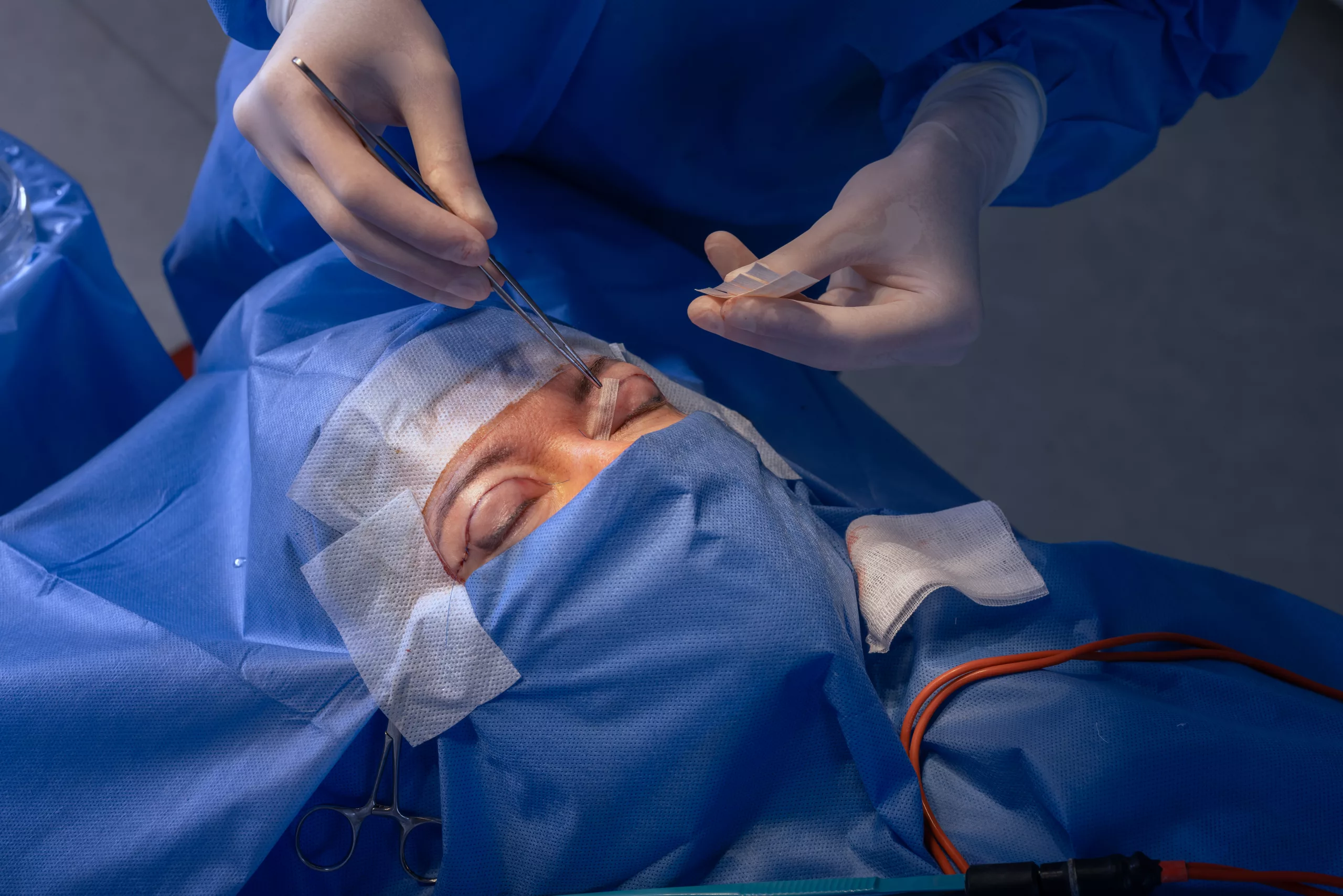
Understanding Transscleral Suture Fixation
Introduction
Transscleral suture fixation is a specialized ophthalmic procedure used to secure intraocular lenses (IOLs) when standard capsular support is unavailable or inadequate. This technique is particularly crucial in complex cataract surgeries and cases where the natural lens or its supporting structures are compromised. In this article, we will delve into the various aspects of transscleral suture fixation, including its indications, procedural details, benefits, potential complications, and post-operative care.
Indications for Transscleral Suture Fixation
Transscleral suture fixation is indicated in scenarios where traditional IOL implantation methods are not viable. These include:
- Aphakia Without Capsular Support: This condition occurs when the eye’s natural lens is absent, and there is no remaining capsular bag to support an IOL.
- Zonular Weakness or Absence: Zonules are the tiny fibers that hold the lens in place. If they are weak or absent due to trauma, disease, or congenital conditions, standard IOL placement becomes challenging.
- Complex Cataract Cases: In cases of cataract surgery complications, such as posterior capsular rupture or dislocation of the lens nucleus, transscleral suture fixation can provide stable IOL placement.
- Previous Vitrectomy: Patients who have undergone vitrectomy (removal of the vitreous gel) may lack sufficient support for a conventional IOL.
Procedural Overview
Transscleral suture fixation involves attaching the IOL to the sclera, the white outer layer of the eyeball, using sutures. The procedure requires meticulous surgical technique and is typically performed by an experienced ophthalmic surgeon. The main steps include:
- Preparation: The patient is given local or general anesthesia. The eye is sterilized and prepared for surgery.
- Incision: A small incision is made in the sclera to create access to the interior of the eye.
- IOL Selection: An appropriate IOL is chosen, considering factors like the patient’s eye anatomy and visual requirements.
- Suture Placement: Sutures, often made of polypropylene or a similar material, are threaded through the sclera and tied securely to the IOL. The number and positioning of sutures depend on the specific case and surgeon’s preference.
- IOL Insertion: The IOL is carefully positioned in the eye, with the sutures ensuring it remains stable.
- Closure: The incision is closed, and the eye is patched or shielded to protect it during the initial healing period.
Benefits of Transscleral Suture Fixation
Transscleral suture fixation offers several advantages, particularly in challenging clinical scenarios:
- Versatility: It provides a solution for patients who cannot benefit from standard IOL implantation due to inadequate capsular support.
- Stability: Properly placed sutures offer robust and long-lasting support for the IOL, reducing the risk of dislocation.
- Improved Visual Outcomes: By ensuring the IOL remains in the correct position, patients can achieve better visual acuity and quality of vision.
- Adaptability: This technique can be tailored to individual patient needs, considering their unique ocular anatomy and medical history.
Potential Complications
Like any surgical procedure, transscleral suture fixation carries some risks. It is essential for patients to be aware of these potential complications and for surgeons to take steps to minimize them. Common complications include:
- Infection: Although rare, there is a risk of infection which can lead to endophthalmitis, a severe eye infection.
- Suture Erosion or Breakage: Over time, sutures may erode through the sclera or break, potentially causing IOL instability.
- Glaucoma: Increased intraocular pressure can occur postoperatively, necessitating careful monitoring and management.
- Retinal Detachment: The procedure can increase the risk of retinal detachment, particularly in patients with pre-existing retinal conditions.
- Inflammation: Post-operative inflammation can affect recovery and visual outcomes, requiring anti-inflammatory medications.
Postoperative Care and Recovery
Post-operative care is crucial for ensuring a successful outcome following transscleral suture fixation. Patients should follow their surgeon’s instructions closely, which typically include:
- Medications: Use prescribed antibiotic and anti-inflammatory eye drops to prevent infection and control inflammation.
- Follow-Up Visits: Attend all scheduled follow-up appointments to monitor healing and detect any early signs of complications.
- Activity Restrictions: Avoid strenuous activities, heavy lifting, and eye rubbing during the initial healing period.
- Protective Eyewear: Use protective eyewear, especially during sleep, to prevent accidental trauma to the operated eye.
- Report Symptoms: Promptly report any unusual symptoms, such as pain, vision changes, or increased redness, to the surgeon.
Conclusion
Transscleral suture fixation is a valuable surgical technique in the realm of ophthalmology, offering a solution for patients with complex cataract cases and insufficient capsular support. While it comes with certain risks, its benefits in providing stable and improved vision make it a critical option in the surgical management of aphakia and related conditions. With proper surgical technique and diligent post-operative care, patients can achieve favorable outcomes and enhanced quality of life.
World Eye Care Foundation’s eyecare.live brings you the latest information from various industry sources and experts in eye health and vision care. Please consult with your eye care provider for more general information and specific eye conditions. We do not provide any medical advice, suggestions or recommendations in any health conditions.
Commonly Asked Questions
Surgeons often use three-piece IOLs or specially designed suturable IOLs due to their stability and ease of fixation.
The surgery typically takes about 1 to 2 hours, depending on the complexity of the case.
Yes, this procedure can be performed on patients of all age groups, including children and elderly individuals, depending on their specific ocular conditions.
Both local anesthesia (with sedation) and general anesthesia can be used, based on the patient’s health status and the surgeon’s preference.
In cases where surgery is not preferred, contact lenses or aphakic spectacles can be used as temporary solutions, though they may not provide the same visual outcomes.
Most patients can resume light activities within a few days, but strenuous activities should be avoided for at least a month post-surgery.
The success rate is generally high, with most patients experiencing significant improvement in vision. However, success depends on individual patient factors and surgical expertise.
It is uncommon to perform the procedure on both eyes simultaneously due to the need for careful post-operative monitoring and recovery.
Sutures can last for many years, but they may require monitoring and potential replacement if they erode or break.
Suture material used in transscleral fixation is generally biocompatible, but in rare cases, patients may develop allergic reactions.
news via inbox
Subscribe here to get latest updates !


