
Comprehensive Guide to Slit-Lamp Biomicroscopy
Introduction
Slit-lamp biomicroscopy is an essential diagnostic procedure in ophthalmology and optometry, offering a detailed and magnified view of the eye’s structures. This advanced technique is critical for the early detection and management of a wide range of ocular conditions, ensuring that eye care professionals can provide the most effective treatments. This comprehensive guide explores the intricacies of slit-lamp biomicroscopy, its components, applications, and significance in ocular health.
What is Slit-Lamp Biomicroscopy?
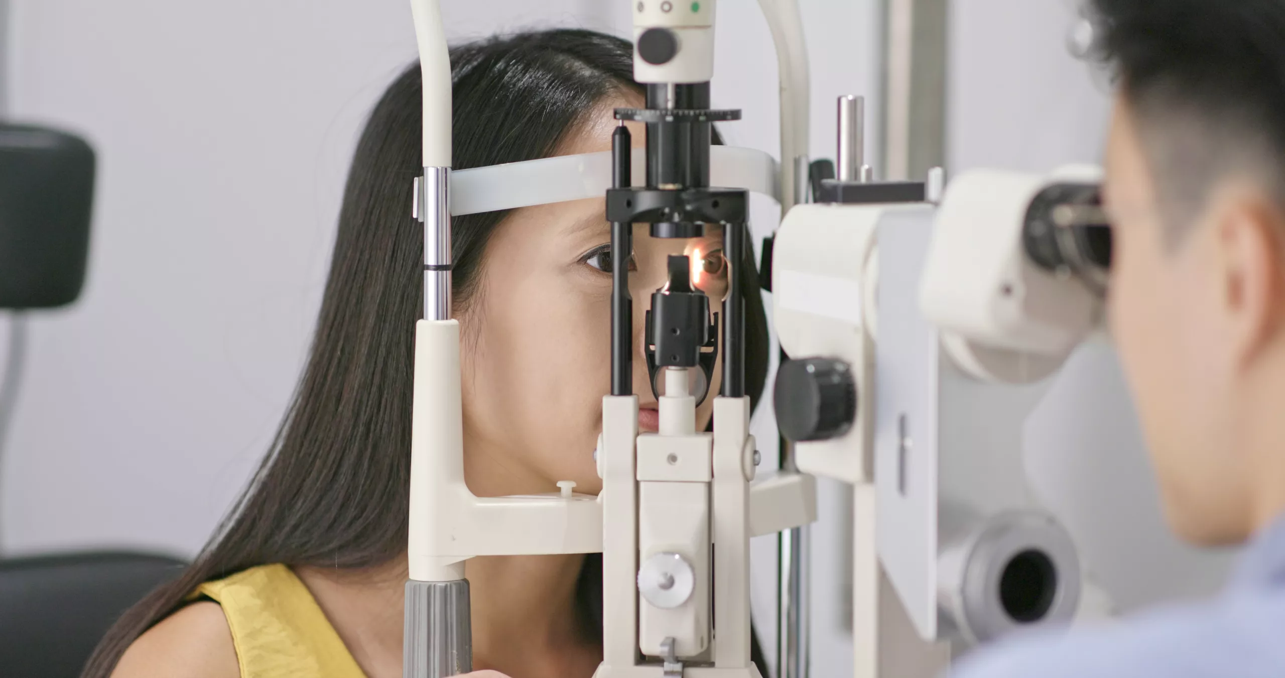
Slit-lamp biomicroscopy involves using a specialized microscope called a slit lamp to examine the eye in detail. This instrument combines a high-intensity light source with a binocular microscope, allowing for the precise visualization of the anterior and posterior segments of the eye under variable magnifications. The slit lamp’s ability to focus on specific eye structures and highlight them with a narrow, slit-like beam of light is what makes it indispensable in eye care.
Components of a Slit Lamp
Understanding the components of a slit lamp helps appreciate its functionality:
- Base and Arm: The base provides stability, while the arm allows for precise movement and positioning of the microscope and light source.
- Illumination System: This includes a high-intensity light source, slit aperture, and various filters. The light beam’s width, height, and angle can be adjusted to enhance the visualization of different eye structures.
- Microscope: The binocular microscope offers stereoscopic (three-dimensional) vision and is equipped with adjustable magnification settings to view eye structures in fine detail.
- Joystick and Control Knobs: These allow the examiner to maneuver the slit lamp smoothly and adjust the focus and alignment during the examination.
- Patient Positioning System: A chin rest and forehead support help stabilize the patient’s head, ensuring accurate and consistent observations.
Procedure
During a slit-lamp examination, the patient is seated comfortably with their chin and forehead placed against the supports to minimize head movement. The examiner uses the joystick and control knobs to adjust the light beam and microscope to focus on specific parts of the eye. Here’s a step-by-step breakdown of the procedure:
- Patient Preparation: The patient is positioned correctly, and the examiner explains the procedure to ensure cooperation.
- Initial Examination: The examiner starts with a broad, low-intensity light to get an overall view of the eye.
- Detailed Observation: By narrowing the light beam and increasing magnification, the examiner can focus on specific areas such as the cornea, lens, and retina.
- Special Techniques: Techniques like retroillumination (shining light through the pupil to illuminate the lens) and specular reflection (using light to view the corneal endothelium) are used for detailed examination.
- Documentation: Findings may be documented using digital imaging systems attached to the slit lamp.
Anterior Segment Examination
- Cornea
- Detecting Corneal Abrasions and Ulcers: The slit lamp can reveal minute corneal defects, helping in diagnosing abrasions, ulcers, and dystrophies.
- Corneal Edema and Opacities: It can detect swelling (edema) and opacities, which may indicate infections, inflammation, or other corneal diseases.
- Keratoconus: The conical deformation and thinning characteristic of keratoconus are easily visible with a slit lamp.
- Conjunctiva and Sclera
- Inflammation and Infection: The slit lamp aids in diagnosing conditions like conjunctivitis (inflammation of the conjunctiva), episcleritis, and scleritis (inflammation of the sclera).
- Foreign Bodies: The detailed view helps locate and assess foreign bodies on the ocular surface.
- Iris and Anterior Chamber
Posterior Segment Examination
- Lens
- Cataracts: The slit lamp is essential for diagnosing cataracts, providing detailed views to determine their type and severity.
- Lens Dislocations: It helps identify dislocations or subluxations of the lens.
- Vitreous and Retina
- Retinal Detachments: Early detection of retinal tears and detachments can prevent vision loss.
- Macular Degeneration: The slit lamp helps assess changes in the macula indicative of age-related macular degeneration (AMD).
- Diabetic Retinopathy: Microaneurysms, hemorrhages, and neovascularization associated with diabetic retinopathy are visible.
Importance in Diagnosing Ocular Conditions
Slit-lamp biomicroscopy is invaluable for diagnosing a wide range of eye conditions, enabling early detection and intervention that can prevent vision loss and improve outcomes.
Common Conditions Diagnosed
- Glaucoma: Assessing the optic nerve head and anterior chamber angle for early signs of glaucoma.
- Dry Eye Syndrome: Evaluating tear film stability and ocular surface abnormalities.
- Conjunctivitis: Identifying bacterial, viral, or allergic conjunctivitis through detailed examination of the conjunctiva.
- Keratitis: Detecting inflammation of the cornea, which could be due to infection or other causes.
Enhancing Diagnostic Accuracy
Advanced techniques and accessories enhance the diagnostic capabilities of slit-lamp biomicroscopy:
- Fluorescein Staining: This involves applying a fluorescent dye to the eye, highlighting corneal abrasions and defects in the tear film.
- Gonioscopy: Specialized lenses allow detailed viewing of the anterior chamber angle, critical for diagnosing types of glaucoma.
- Tonometry: Integrated tonometry devices measure intraocular pressure, aiding in glaucoma diagnosis.
- Imaging Systems: Digital cameras and video systems attached to the slit lamp facilitate documentation and analysis of findings, enabling better monitoring and sharing of information.
Training and Expertise
Proper training is essential for practitioners to effectively use the slit lamp. Training involves:
- Understanding Equipment: Knowing the functions and settings of the slit lamp components.
- Mastering Techniques: Learning various examination techniques, such as diffuse illumination, retroillumination, and specular reflection.
- Staying Updated: Keeping abreast of advancements in slit-lamp technology and ocular diagnostics.
Conclusion
Slit-lamp biomicroscopy is a cornerstone of modern ocular diagnostics, offering unparalleled insights into the eye’s health. Its comprehensive capabilities make it indispensable for diagnosing and managing a wide array of eye conditions. By enhancing diagnostic accuracy and enabling early intervention, slit-lamp biomicroscopy plays a vital role in preserving and improving vision health.
World Eye Care Foundation’s eyecare.live brings you the latest information from various industry sources and experts in eye health and vision care. Please consult with your eye care provider for more general information and specific eye conditions. We do not provide any medical advice, suggestions or recommendations in any health conditions.
Commonly Asked Questions
Slit-lamp biomicroscopy is used to examine the anterior and posterior segments of the eye in detail, aiding in the diagnosis and management of various eye conditions such as cataracts, glaucoma, and diabetic retinopathy.
A slit lamp combines a high-intensity light source with a binocular microscope, allowing eye structures to be viewed under magnification. It uses a narrow slit beam to illuminate specific areas like the cornea, lens, and retina.
Components include a base and arm for stability and movement, an illumination system with adjustable light beam settings, a binocular microscope for stereoscopic vision, and control knobs for maneuvering the instrument.
Techniques include retroillumination for viewing the lens and specular reflection for examining the corneal endothelium, enhancing detailed observation of ocular structures.
It allows for the assessment of the optic nerve head and anterior chamber angle, crucial in detecting early signs of glaucoma and monitoring disease progression.
Yes, it can visualize microaneurysms, hemorrhages, and neovascularization in the retina associated with diabetic retinopathy, facilitating early intervention to prevent vision loss.
It provides detailed views of the lens to diagnose different types and stages of cataracts, guiding treatment decisions such as surgical intervention when necessary.
It can detect corneal abrasions, ulcers, edema, and opacities, which are indicative of infections, inflammation, or other conditions affecting the cornea.
Proper training ensures eye care professionals can effectively operate the slit lamp, master examination techniques, and stay updated with advancements in ocular diagnostics.
Imaging systems attached to the slit lamp enable digital documentation and analysis of findings, improving diagnostic accuracy, and facilitating collaboration among healthcare providers.


news via inbox
Subscribe here to get latest updates !


