
Unveiling Pseudoexfoliation Syndrome
Introduction
Pseudoexfoliation syndrome (PXF) is a significant age-related condition that affects the eye’s structure, leading to complications such as glaucoma. This detailed guide explores the intricacies of PXF, its symptoms, diagnostic processes, treatment options, and preventive measures. Our goal is to provide comprehensive insights to enhance understanding and management of this condition.
What is Pseudoexfoliation Syndrome?
Pseudoexfoliation syndrome is a progressive disorder characterized by the accumulation of abnormal fibrillar material on the eye’s lens, iris, and trabecular meshwork. This material, composed primarily of extracellular matrix proteins, interferes with the eye’s drainage system, leading to increased intraocular pressure (IOP) and potential vision loss.
Pathophysiology and Causes
- Genetic Factors: The exact etiology of PXF remains elusive, though several factors are believed to contribute:
- LOXL1 Gene Mutation: The LOXL1 gene mutation is strongly associated with PXF. This gene is crucial for the synthesis of elastin and collagen, influencing the structural integrity of ocular tissues. Variants of the LOXL1 gene have been shown to increase susceptibility to PXF. However, not everyone with these gene variants develops PXF, suggesting other genetic or environmental factors play a role.
- Other Genetic Factors: Research indicates that other genes might also be involved, contributing to the complexity of the disease’s inheritance patterns.
- Environmental Factors
- Ultraviolet (UV) Exposure: Chronic exposure to UV radiation is thought to contribute to the development of PXF. UV light may induce oxidative stress, which damages ocular tissues and promotes the accumulation of pseudoexfoliative material.
- Geographic Location: PXF is more prevalent in certain regions, suggesting that geographic and climatic factors may influence its development. Populations in northern latitudes with high UV exposure, such as Scandinavia, show a higher prevalence.
- Lifestyle and Diet: Factors such as diet, smoking, and alcohol consumption have been studied, but their roles in PXF development remain unclear. A diet rich in antioxidants may help mitigate some of the oxidative stress associated with PXF.
- Age
- Incidence by Age: PXF predominantly affects individuals over the age of 60, with its incidence increasing with age. The disease is rare in younger individuals, suggesting that age-related changes in the eye’s extracellular matrix contribute to its development.
Symptoms of Pseudoexfoliation Syndrome
PXF is often asymptomatic in its early stages. As the disease progresses, patients may experience:
- Elevated Intraocular Pressure (IOP)
- Significance of Elevated IOP: This is the most significant risk factor for glaucoma. Elevated IOP can damage the optic nerve, leading to irreversible vision loss. It occurs due to the accumulation of pseudoexfoliative material in the trabecular meshwork, obstructing aqueous humor outflow.
- Vision Disturbances
- Blurred Vision: Patients may notice blurriness or fluctuating vision due to the deposition of pseudoexfoliative material on the lens, causing lens opacity and cataract formation. This material can also cause corneal endothelial dysfunction, contributing to visual disturbances.
- Iris Abnormalities
- Atrophy and Transillumination Defects: The iris may exhibit signs of atrophy, transillumination defects, or changes in pigmentation. This can be observed as areas where light shines through the iris due to loss of pigment cells.
- Lens Changes
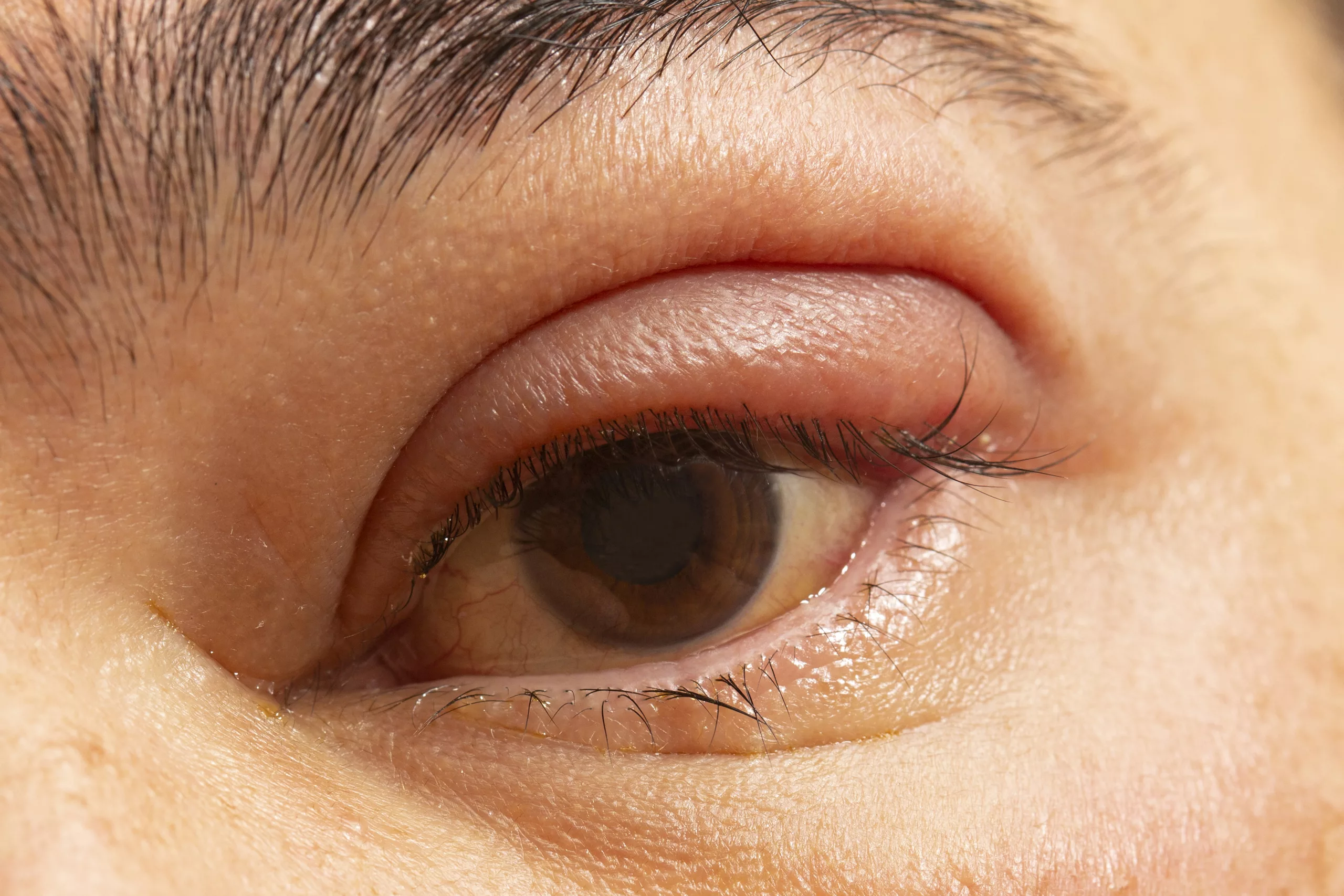
- Pseudoexfoliative Material Deposits: Deposits of pseudoexfoliative material on the anterior lens capsule are often visible during a slit-lamp examination. These changes can lead to cataract formation, complicating surgical interventions. The material often forms a characteristic three-ring pattern on the lens capsule.
Comprehensive Diagnosis of Pseudoexfoliation Syndrome
Diagnosing PXF involves a detailed ocular examination and the use of advanced diagnostic tools:
- Clinical Examination
- Slit-Lamp Biomicroscopy
- Detection of Pseudoexfoliative Material: This is the cornerstone for diagnosing PXF. The presence of white, flaky material on the anterior lens capsule is a hallmark sign. The material can also be found on the corneal endothelium, pupillary border, and other anterior segment structures.
- Gonioscopy
- Assessment of the Trabecular Meshwork: Essential for assessing the trabecular meshwork and identifying pseudoexfoliative material in the angle, which is critical for glaucoma diagnosis. Gonioscopy helps to visualize the drainage angle and detect any angle closure or abnormalities.
- Slit-Lamp Biomicroscopy
- Tonometry
- Measurement of IOP: Measurement of IOP is vital. Elevated IOP is a significant indicator of glaucoma risk associated with PXF. Regular IOP monitoring is crucial for early intervention and management.
- Advanced Diagnostic Techniques
- Optical Coherence Tomography (OCT)
- High-Resolution Imaging: OCT provides high-resolution images of the retina and optic nerve head, helping to detect early glaucomatous changes. It can measure retinal nerve fiber layer thickness and ganglion cell complex, which are vital in glaucoma diagnosis and monitoring.
- Optical Coherence Tomography (OCT)
- Fundus Photography and Imaging
- Visualization of the Optic Nerve Head: These techniques are used to visualize the optic nerve head and retinal nerve fiber layer, assessing for signs of glaucoma. Serial photographs can help track changes over time.
- Ultrasound Biomicroscopy (UBM)
- Evaluation of Anterior Segment Structures: This imaging modality is particularly useful for evaluating the anterior segment structures, including the angle and ciliary body. UBM provides detailed images of the ciliary processes, which can be affected in PXF.
Treatment Options for Pseudoexfoliation Syndrome
Managing PXF focuses on controlling IOP to prevent glaucomatous damage. Treatment modalities include:
Medications
- Topical Eye Drops
- Prostaglandin Analogues: Increase aqueous outflow through the uveoscleral pathway. Common drugs include latanoprost, bimatoprost, and travoprost.
- Beta-Blockers: Reduce aqueous humor production. Commonly used beta-blockers include timolol and betaxolol.
- Alpha Agonists: Decrease aqueous production and increase outflow. Examples include brimonidine and apraclonidine.
- Carbonic Anhydrase Inhibitors: Reduce aqueous humor production, often used in combination therapy. Dorzolamide and brinzolamide are examples of topical carbonic anhydrase inhibitors.
- Oral Medications
- Carbonic Anhydrase Inhibitors: Oral medications like acetazolamide and methazolamide may be prescribed to lower IOP when topical treatments are insufficient. These drugs can have systemic side effects and are usually reserved for short-term use or severe cases.
Laser Treatments
- Laser Trabeculoplasty
- Selective Laser Trabeculoplasty (SLT): SLT uses laser energy to target specific cells in the trabecular meshwork, enhancing aqueous outflow and lowering IOP. It is often preferred due to its lower risk of complications compared to argon laser trabeculoplasty (ALT).
- Argon Laser Trabeculoplasty (ALT): ALT uses a thermal laser to treat the trabecular meshwork. It is less commonly used now due to higher risks of scarring and complications.
- Laser Cataract Surgery
- Addressing Coexisting Cataract: In cases of coexisting cataract, laser-assisted cataract surgery can be performed, addressing both cataract and PXF-related complications. The use of femtosecond lasers can make the procedure more precise and safer.
Surgical Interventions
- Trabeculectomy
- Creating a New Drainage Pathway: This surgical procedure creates a new drainage pathway for aqueous humor, significantly lowering IOP. It involves creating a small flap in the sclera and a reservoir (bleb) under the conjunctiva where fluid can drain.
- Success Rates and Complications: Trabeculectomy has a high success rate but can be associated with complications such as infection, bleb failure, or hypotony (excessively low IOP).
- Glaucoma Drainage Devices
- Implants for Aqueous Outflow: Implants like Ahmed valves or Baerveldt shunts help manage IOP by facilitating aqueous outflow through a silicone tube. These devices are often used in refractory glaucoma cases where other treatments have failed.
- Combined Cataract and Glaucoma Surgery
- Phacoemulsification and Trabeculectomy: This approach addresses cataract extraction and glaucoma treatment simultaneously, using techniques such as phacoemulsification with a trabeculectomy or implantation of a drainage device. Combined surgery can be more efficient and reduce the need for multiple surgical procedures.
Preventive Strategies and Lifestyle Adjustments
Regular monitoring and proactive management are crucial in preventing complications associated with PXF:
- Regular Eye Examinations
- Annual Check-Ups: Annual comprehensive eye exams are essential for early detection of PXF and glaucoma. Patients should be vigilant about their eye health, especially if they have a family history of the condition.
- Monitoring IOP and Optic Nerve Health: Regular IOP measurements and optic nerve assessments are critical for early intervention and effective management.
- UV Protection
- Sunglasses with UV Protection: Wearing sunglasses with UV protection can help mitigate the risk of PXF progression. Broad-spectrum sunglasses that block 100% of UV rays are recommended.
- Dietary Considerations
- Antioxidant-Rich Diet: A diet rich in antioxidants and omega-3 fatty acids may help reduce inflammation and oxidative stress, potentially slowing disease progression. Foods like leafy greens, fish, nuts, and fruits are beneficial.
- Lifestyle Adjustments
- Avoiding Smoking and Excessive Alcohol: Smoking and excessive alcohol consumption can contribute to oxidative stress and inflammation, potentially exacerbating PXF.
- Regular Physical Activity: Exercise can improve overall health and may help reduce IOP in some individuals.
Conclusion
Pseudoexfoliation syndrome is a multifaceted condition requiring a thorough understanding of its pathophysiology, symptoms, diagnostic techniques, and treatment strategies. By staying informed and adhering to recommended treatment protocols, individuals with PXF can effectively manage their condition and preserve their vision. Regular consultations with an ophthalmologist are vital for adapting the treatment plan to the patient’s evolving needs, ensuring the best possible outcomes for ocular health.
World Eye Care Foundation’s eyecare.live brings you the latest information from various industry sources and experts in eye health and vision care. Please consult with your eye care provider for more general information and specific eye conditions. We do not provide any medical advice, suggestions or recommendations in any health conditions.
Commonly Asked Questions
Pseudoexfoliation Syndrome (PXF) involves the accumulation of abnormal fibrillar material in the eye, which is distinct from true exfoliation syndrome seen in systemic diseases. True exfoliation involves actual peeling of the lens capsule, which is different from the fibrillar deposits seen in PXF.
Yes, pseudoexfoliation syndrome (PXF) can occur in both eyes, but it often starts in one eye before potentially affecting the other. Regular eye exams are crucial for monitoring both eyes if PXF is diagnosed.
PXF has a genetic component, and family history can increase the risk of developing the condition. Mutations in the LOXL1 gene are particularly associated with PXF, but not all carriers of the mutation will develop the syndrome.
PXF leads to the accumulation of exfoliative material in the trabecular meshwork, the drainage system of the eye, causing increased intraocular pressure (IOP). Elevated IOP can damage the optic nerve, leading to glaucoma.
Patients with PXF may face complications such as lens capsule fragility, zonular weakness, and increased risk of intraoperative complications. Surgeons need to take special precautions to manage these risks effectively.
Individuals with PXF should see an ophthalmologist at least once a year. If they have elevated IOP or glaucoma, more frequent visits, possibly every 3-6 months, may be necessary to monitor and manage their condition.
While there are no alternative treatments that cure PXF, complementary approaches like maintaining a healthy diet, using herbal supplements with antioxidant properties, and managing overall health can support conventional treatments.
Surgical risks include complications from cataract surgery, such as zonular weakness leading to lens dislocation, increased risk of postoperative inflammation, and difficulties in managing IOP after surgery.
PXF has been associated with systemic vascular diseases, including cardiovascular issues, but the exact relationship is still under investigation. Patients with PXF may have a higher risk of systemic vascular abnormalities.
While treatments can manage the symptoms and control IOP, PXF is a chronic condition, and the underlying deposition of material can continue, necessitating ongoing management and monitoring.


news via inbox
Subscribe here to get latest updates !


