Advancements in Optical Coherence Tomography (OCT) Technology
Introduction
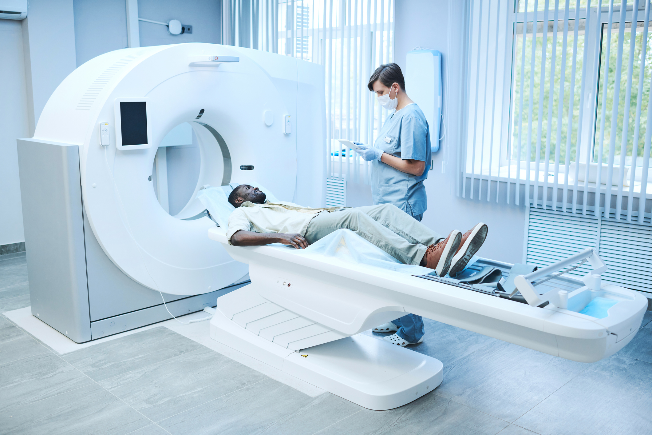
Optical Coherence Tomography (OCT) has emerged as a cornerstone technology in the field of ophthalmology, offering non-invasive, high-resolution imaging of ocular structures. Over the years, OCT has undergone significant advancements, expanding its capabilities beyond mere structural visualization to encompass functional assessments and automated analysis. In this article, we delve into the latest innovations in OCT technology and their transformative impact on ocular imaging and patient care.
Enhanced Resolution and Speed
Modern OCT systems boast remarkable improvements in resolution and scanning speed, enabling clinicians to capture detailed, three-dimensional images of ocular tissues in seconds. Enhanced resolution allows for the visualization of microstructures within the retina, such as individual retinal layers and photoreceptor integrity, aiding in the early detection and monitoring of retinal diseases like macular degeneration and diabetic retinopathy. Moreover, rapid scanning speeds minimize motion artifacts, ensuring high-quality imaging even in patients unable to maintain steady fixation.
Angiography and Microvascular Imaging
OCT angiography (OCTA) represents a significant advancement in functional imaging, providing detailed visualization of retinal and choroidal vasculature without the need for contrast agents. By leveraging motion contrast imaging techniques, OCTA enables the assessment of blood flow dynamics and microvascular perfusion in various retinal pathologies. This includes the detection of neovascularization in conditions like diabetic retinopathy and the evaluation of vascular changes in diseases such as glaucoma and retinal vein occlusions. OCTA holds promise for early disease detection, monitoring treatment response, and guiding therapeutic interventions.
Artificial Intelligence and Machine Learning Integration
The integration of artificial intelligence (AI) and machine learning algorithms with OCT data has revolutionized image analysis and diagnostic decision-making. AI-based software can automatically segment retinal layers, detect subtle changes in tissue morphology, and classify disease severity with high accuracy. Furthermore, machine learning algorithms trained on large datasets can predict disease progression and treatment outcomes, assisting clinicians in personalized treatment planning and prognostication. These AI-driven tools streamline workflow, enhance diagnostic efficiency, and improve patient outcomes by facilitating timely interventions.
Handheld and Portable OCT Devices
The development of handheld and portable OCT devices has democratized access to advanced imaging technology, particularly in underserved or remote regions. These compact systems offer real-time visualization of ocular structures outside traditional clinical settings, facilitating point-of-care diagnostics and monitoring. Portable OCT devices play a crucial role in community outreach programs, telemedicine initiatives, and emergency triage situations, where immediate assessment of ocular health is paramount. By bringing OCT to the bedside, these devices empower healthcare providers to deliver timely interventions and improve patient outcomes, especially in resource-limited settings.
Conclusion
Advancements in Optical Coherence Tomography (OCT) technology have propelled the field of ocular imaging into a new era of precision medicine and personalized care. From enhanced resolution and functional angiography to AI-driven analysis and portable devices, these innovations are reshaping the landscape of ophthalmic diagnostics and therapeutics. As OCT continues to evolve, its integration with other imaging modalities and data analytics tools holds promise for further enhancing our understanding of ocular diseases and optimizing patient management strategies. By leveraging the power of OCT technology, clinicians can achieve earlier disease detection, more accurate diagnosis, and tailored treatment approaches, ultimately improving visual outcomes and quality of life for patients worldwide.
World Eye Care Foundation’s eyecare.live brings you the latest information from various industry sources and experts in eye health and vision care. Please consult with your eye care provider for more general information and specific eye conditions. We do not provide any medical advice, suggestions or recommendations in any health conditions.
Commonly Asked Questions
Future advancements in OCT technology may include further improvements in resolution and speed, integration with other imaging modalities, and enhanced AI-driven analysis for predictive modeling and treatment optimization.
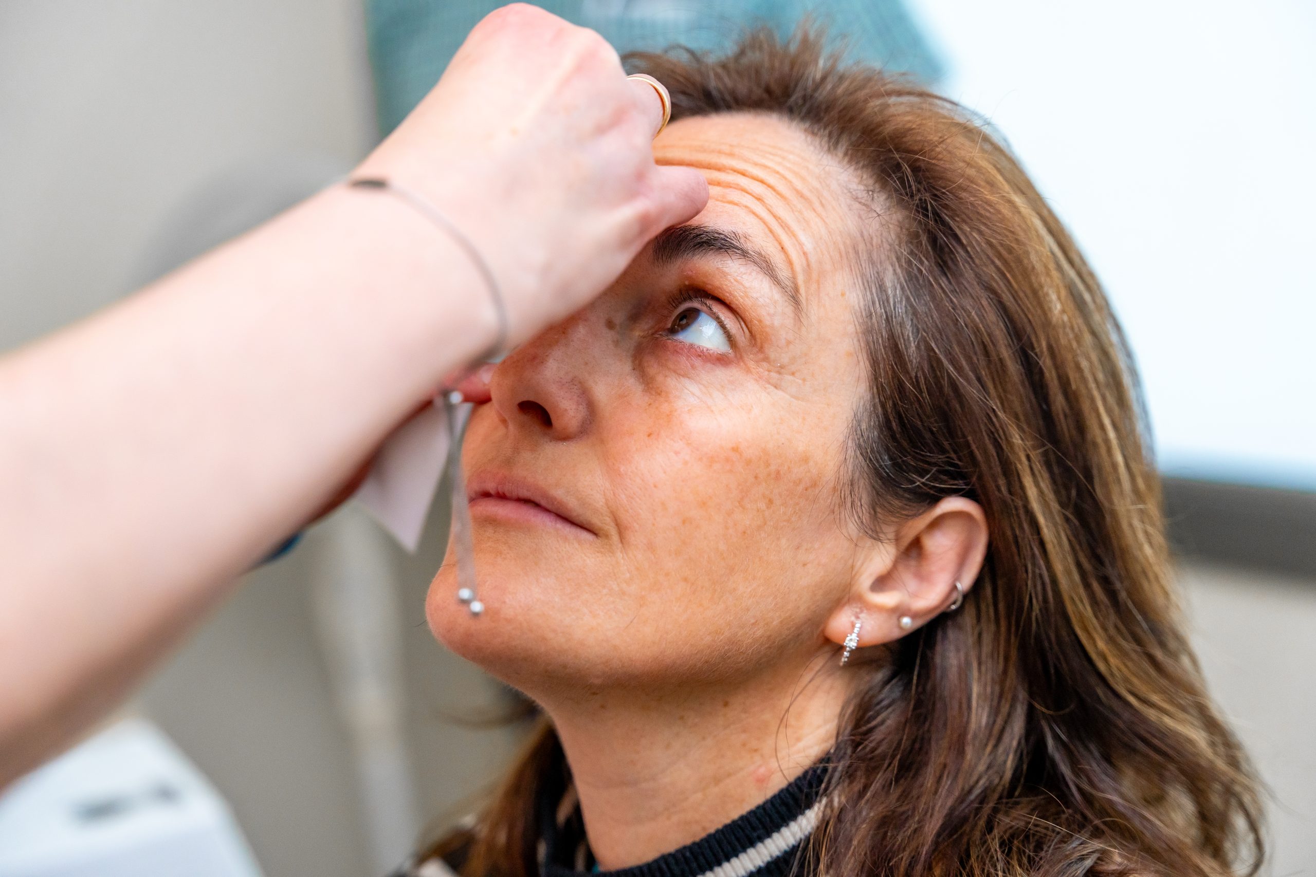
OCT imaging is non-invasive and generally safe. However, patients may experience mild discomfort from pupil dilation or eye drops used during the examination. Rarely, allergic reactions to imaging agents may occur, but this is uncommon.
The frequency of OCT imaging depends on the underlying retinal condition and the treatment regimen. In many cases, OCT scans are performed at regular intervals to monitor disease progression, treatment response, and therapeutic efficacy.
Portable OCT devices complement traditional imaging systems by offering point-of-care diagnostics in remote or resource-limited settings. While they may not replace conventional systems, they expand access to advanced imaging technology and facilitate timely interventions.
AI algorithms are used to automate image segmentation, detect pathology, and classify disease severity based on OCT data. This technology enhances diagnostic accuracy, streamlines workflow, and assists in personalized treatment planning.
While OCT provides excellent visualization of retinal structures, it may have limitations in imaging through media opacities such as cataracts or vitreous hemorrhage. Additionally, motion artifacts can affect image quality, particularly in patients unable to maintain steady fixation.
Yes, OCT imaging can detect structural changes associated with glaucoma, such as thinning of the retinal nerve fiber layer and optic nerve head cupping. These changes can be indicative of early glaucomatous damage.
Unlike traditional dye-based angiography, OCT angiography provides non-invasive visualization of retinal and choroidal vasculature, without the need for contrast agents. It offers detailed assessment of blood flow dynamics and microvascular perfusion.
OCT is used for diagnosing and managing various ocular conditions, including macular degeneration, diabetic retinopathy, glaucoma, and retinal vascular diseases.
OCT is a non-invasive imaging technique used to visualize the microstructures of the eye. It works by measuring the echo time delay and intensity of light reflected from ocular tissues, producing high-resolution cross-sectional images.
news via inbox
Subscribe here to get latest updates !


