Angioid Streaks Unveiled: Causes, Symptoms, and Management
Angioid Streaks are a rare eye condition characterized by breaks in the Bruch’s membrane. This article aims to provide clarity on the causes, symptoms, and eye care guidelines for Angioid Streaks. Learn when to seek medical attention, potential complications, risk factors, preventive measures, diagnosis methods, treatment options, and insights for optimal eye health in individuals dealing with this condition.
Overview of Angioid Streaks
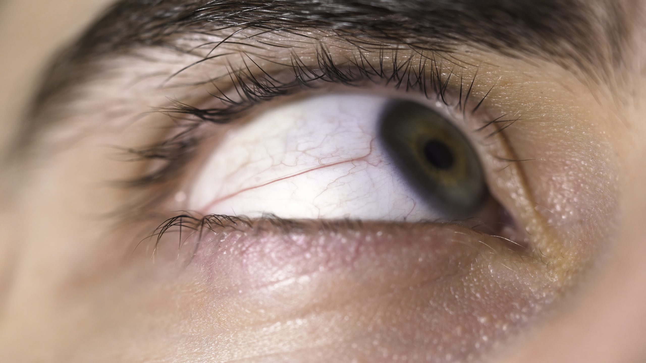
Angioid streaks are visible breaks or cracks in Bruch’s membrane, a layer of tissue located beneath the retina in the eye. Bruch’s membrane plays a crucial role in supporting and nourishing the retina. Angioid streaks appear as dark lines or irregular streaks on the fundus, which is the back of the eye that can be seen during an eye examination.
Symptoms
- Distorted Vision: Angioid streaks can lead to distorted or blurry vision, especially if they involve the central part of the retina (macula).
- Metamorphopsia: Some individuals may experience metamorphopsia, a condition where straight lines appear wavy or distorted.
- Reduced Visual Acuity: In advanced cases, angioid streaks may contribute to a gradual reduction in visual acuity.
- Central Scotomas: Blind spots or central scotomas may occur, impacting the central field of vision.
Causes
- Elastin Degradation: The primary cause is the degeneration of elastin, a protein that provides elasticity to tissues, including Bruch’s membrane.
- Underlying Conditions: Angioid streaks are often associated with systemic conditions such as pseudoxanthoma elasticum (PXE), Ehlers-Danlos syndrome, or Paget’s disease.
- Trauma: Severe eye trauma, such as a blunt injury, can lead to the development of angioid streaks.
What Happens Because of the Condition
- Breaks in Bruch’s Membrane: Angioid streaks represent breaks or fissures in Bruch’s membrane, compromising its integrity.
- Choroidal Neovascularization: In some cases, angioid streaks can lead to the growth of abnormal blood vessels from the choroid into the retina, a condition known as choroidal neovascularization (CNV).
- Retinal Detachment: In severe cases, angioid streaks may contribute to retinal detachment, leading to a sudden and significant loss of vision.
Risk Factors
- Systemic Conditions: Individuals with systemic conditions such as pseudoxanthoma elasticum (PXE), Ehlers-Danlos syndrome, or Paget’s disease are at an increased risk.
- Genetic Factors: There may be a genetic predisposition, with a family history of angioid streaks increasing the likelihood of development.
- Trauma: Severe eye trauma, even if it occurred years before, is a potential risk factor.
Diagnosis
- Fundus Examination: An ophthalmologist will perform a fundus examination using ophthalmoscopy to visualize the back of the eye and identify angioid streaks.
- Fluorescein Angiography: This imaging test involves injecting a dye into the bloodstream, which highlights the blood vessels in the retina. It helps identify the extent of choroidal neovascularization.
- Optical Coherence Tomography (OCT): OCT provides detailed cross-sectional images of the retina, assisting in assessing the structural changes associated with angioid streaks.
- Medical History: Gathering a comprehensive medical history, including information about systemic conditions and any history of eye trauma, is crucial for accurate diagnosis.
Treatment Options
- Observation: In some cases, particularly when angioid streaks are asymptomatic and not causing significant visual impairment, a strategy of close observation may be employed. Regular eye exams to monitor any changes are crucial.
- Anti-VEGF Therapy: If choroidal neovascularization (CNV) is present and contributing to visual impairment, intravitreal injections of anti-vascular endothelial growth factor (anti-VEGF) medications may be considered. These drugs aim to inhibit the growth of abnormal blood vessels, helping to manage CNV.
- Photodynamic Therapy (PDT): PDT involves the use of a photosensitive drug and laser light to selectively treat abnormal blood vessels associated with CNV. This therapy is sometimes used in combination with anti-VEGF treatment.
- Subretinal Surgery: In cases of retinal detachment caused by angioid streaks, surgical interventions such as subretinal surgery may be considered to reattach the retina and restore visual function.
Complications
- Choroidal Neovascularization (CNV): CNV is a significant complication that can lead to vision loss. Early detection and treatment with anti-VEGF therapy or PDT can help manage this complication.
- Retinal Detachment: Angioid streaks increase the risk of retinal detachment. Surgical interventions may be necessary to address retinal detachment and restore vision.
- Visual Impairment: The progression of angioid streaks can result in a gradual decline in visual acuity, impacting daily activities. Timely management is essential to mitigate vision loss.
Prevention
- Manage Systemic Conditions: For individuals with underlying systemic conditions associated with angioid streaks, such as pseudoxanthoma elasticum (PXE), proactive management and treatment of these conditions may help reduce the risk.
- Regular Eye Examinations: Routine eye examinations, especially for individuals with a family history of angioid streaks or other predisposing factors, can facilitate early detection and intervention.
- Protective Eyewear: Individuals engaged in activities with an increased risk of eye trauma, such as certain sports or occupations, should wear appropriate protective eyewear to minimize the risk of injuries that could contribute to angioid streaks.
Medications
Anti-VEGF Medications: Drugs such as ranibizumab or aflibercept may be prescribed to inhibit vascular endothelial growth factor, which plays a role in the growth of abnormal blood vessels. These medications are administered through intravitreal injections.
When to See a Doctor
- Visual Symptoms: Individuals experiencing visual symptoms such as distorted or blurry vision, metamorphopsia (distorted perception of straight lines), or central scotomas (blind spots) should seek prompt evaluation by an eye care professional.
- History of Systemic Conditions: Individuals with a known history of systemic conditions associated with angioid streaks, such as pseudoxanthoma elasticum (PXE), should undergo regular eye examinations, and any new visual symptoms warrant a visit to the doctor.
- Changes in Vision: Any sudden or gradual changes in vision, regardless of age, should be promptly addressed by an eye care professional. This includes a decline in visual acuity, changes in color perception, or any other visual disturbances.
- Family History: Individuals with a family history of angioid streaks or related systemic conditions should be vigilant about regular eye check-ups, and any symptoms should prompt a visit to the doctor.
Demographics More Susceptible
- Systemic Conditions: Individuals with systemic conditions associated with angioid streaks, such as pseudoxanthoma elasticum (PXE), Ehlers-Danlos syndrome, or Paget’s disease, are more susceptible. These individuals should be proactive in seeking regular eye examinations.
- Age: While angioid streaks can occur at any age, they are often diagnosed in adulthood. Adults, especially those over the age of 40, should be mindful of changes in vision and seek timely evaluation.
Follow-up Care for Adults and Children
Follow-up Care for Children:
- Pediatric Ophthalmologist Visits: Children diagnosed with angioid streaks, especially in the context of associated systemic conditions, may require ongoing visits to a pediatric ophthalmologist for regular eye examinations.
- Monitoring Visual Development: Regular assessments of visual development and acuity are essential to identify any changes and implement appropriate interventions.
Follow-up Care for Adults:
- Ophthalmologist Visits: Adults with angioid streaks should continue to see an ophthalmologist for periodic eye examinations. The frequency of visits will depend on the severity of the condition and the presence of complications.
- Choroidal Neovascularization Monitoring: For individuals with choroidal neovascularization (CNV), regular monitoring is crucial. This may involve imaging studies such as fluorescein angiography or optical coherence tomography (OCT) to assess the activity of abnormal blood vessels.
Conclusion
In conclusion, individuals experiencing visual symptoms, especially those with a history of systemic conditions associated with angioid streaks, should promptly consult an eye care professional. Regular eye examinations, particularly for those in high-risk demographics, are essential for early detection and intervention. Children with angioid streaks may need ongoing monitoring by a pediatric ophthalmologist to ensure optimal visual development. Adults should continue to see an ophthalmologist for periodic evaluations, especially if complications such as choroidal neovascularization are present. A proactive approach to eye health, timely visits to eye care professionals, and adherence to recommended follow-up care contribute to the overall well-being and preservation of vision for individuals with angioid streaks.
World Eye Care Foundation’s eyecare.live brings you the latest information from various industry sources and experts in eye health and vision care. Please consult with your eye care provider for more general information and specific eye conditions. We do not provide any medical advice, suggestions or recommendations in any health conditions.
Commonly Asked Questions
The progression of angioid streaks varies among individuals, and regular monitoring is crucial to address any changes.
Laser treatment is not typically used for angioid streaks, but anti-VEGF injections may be considered in certain cases.
Preventive measures involve managing underlying conditions and maintaining regular eye check-ups for early detection.
It can affect individuals of any ethnicity, but prevalence may vary based on underlying conditions.
Angioid streaks do not resolve on their own, and professional management is necessary.
Consultation with an eye specialist is recommended to determine the suitability of contact lenses based on individual circumstances.
In some cases, there may be a genetic component, especially if associated with conditions like pseudoxanthoma elasticum.
Angioid streaks are not visible on the surface but can be detected through comprehensive eye examinations and imaging tests.
There is no specific cure, but management involves addressing underlying conditions and preventing complications.
While angioid streaks themselves may not cause blindness, complications like choroidal neovascularization can lead to vision loss.


news via inbox
Subscribe here to get latest updates !