Blepharophimosis Syndrome Demystified: What You Need to Know
Blepharophimosis Syndrome is a congenital condition characterized by eyelid abnormalities and other facial features. This article aims to provide clarity on the causes, symptoms, and comprehensive eye care for Blepharophimosis Syndrome. Learn when to seek medical attention, potential complications, risk factors, preventive measures, diagnosis methods, treatment options, and insights for optimal eye health in individuals dealing with this syndrome.
Overview of Blepharophimosis Syndrome
Blepharophimosis syndrome is a rare congenital disorder characterized by a combination of distinctive facial features. It primarily affects the eyelids, resulting in a narrowing of the eye openings (blepharophimosis), but it can also involve other facial structures.
Symptoms
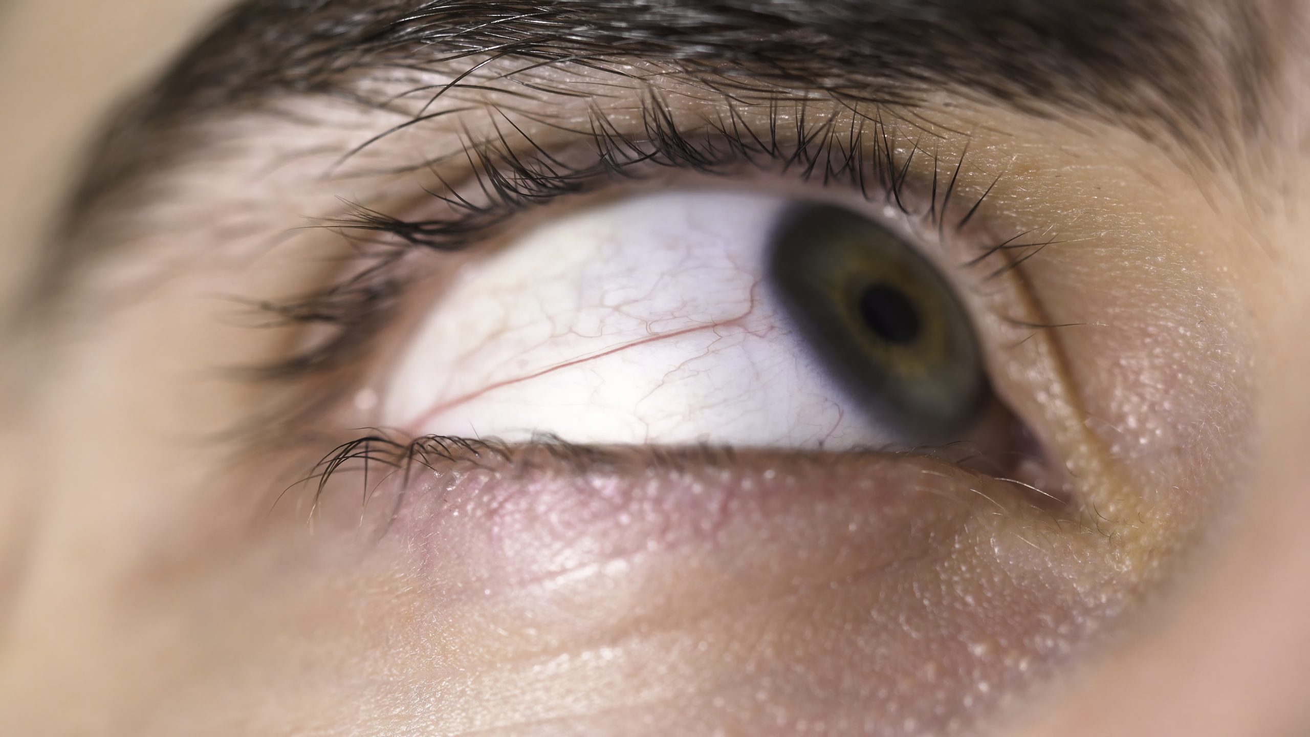
- Narrow Eyelid Opening (Blepharophimosis): The primary feature is a reduced horizontal width of the eyelid opening, leading to a narrow appearance.
- Ptosis: Individuals with blepharophimosis syndrome may experience ptosis, where the upper eyelids droop, partially covering the eyes.
- Epicanthus Inversus: Epicanthus inversus refers to a fold of skin on the inner corner of the eyes, creating a distinctive appearance.
- Telecanthus: Some individuals may exhibit telecanthus, an increased distance between the inner corners of the eyes.
Causes
- Genetic Mutations: Blepharophimosis syndrome is often caused by genetic mutations affecting the development of the eyelids.
- Autosomal Dominant Inheritance: In some cases, the condition may be inherited in an autosomal dominant pattern, meaning one copy of the altered gene from either parent can lead to the syndrome.
What Happens Because of the Condition
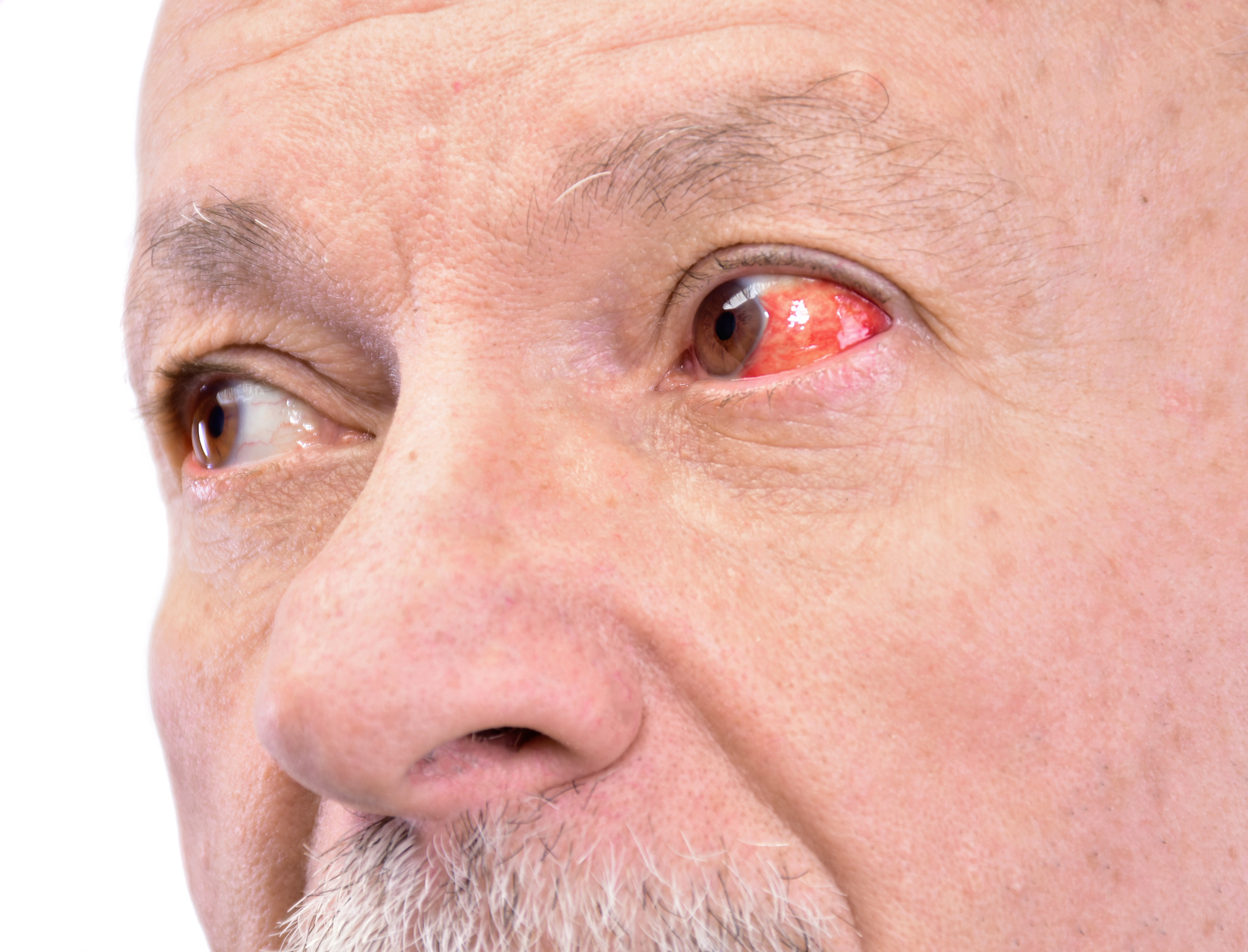
- Visual Impairment: The reduced width of the eyelid opening and ptosis can potentially impact vision by limiting the field of view.
- Aesthetic Concerns: Blepharophimosis syndrome can result in distinctive facial features, leading to aesthetic concerns that may affect an individual’s self-perception.
Risk Factors
- Genetic Predisposition: Individuals with a family history of telecanthus or related craniofacial conditions may have a higher risk.
- Congenital Syndromes: Conditions associated with congenital syndromes, where telecanthus is a characteristic feature, may increase susceptibility. Examples include Down syndrome, Apert syndrome, or other craniofacial disorders.
- Facial Trauma or Injury: Trauma to the facial bones, particularly around the eye area, can contribute to structural changes leading to telecanthus.
- Abnormal Fetal Development: Issues during fetal growth that impact craniofacial development may result in telecanthus.
- Orthodontic or Dental Factors: Malocclusion or orthodontic issues that affect the alignment of the jaw and facial bones can contribute to telecanthus.
Diagnosis
- Physical Examination: A thorough physical examination by a qualified healthcare professional is crucial. This includes measuring the intercanthal distance, which is the distance between the inner corners of the eyes.
- Medical History: Gathering information about any congenital conditions, genetic predisposition, or history of facial trauma is essential for accurate diagnosis.
- Craniofacial Imaging: In some cases, craniofacial imaging such as X-rays, CT scans, or magnetic resonance imaging (MRI) may be recommended. Imaging helps assess the underlying structural factors contributing to telecanthus.
- Ophthalmologic Evaluation: A comprehensive eye examination by an ophthalmologist is important to assess the impact of telecanthus on visual function and eye health.
- Genetic Counseling: If there is a suspicion of genetic factors contributing to telecanthus, genetic counseling may be recommended, especially for individuals with a family history or those with congenital syndromes.
Treatment Options
- Surgical Correction: Depending on the underlying cause and severity, surgical interventions may be considered. Procedures aim to reposition the inner corners of the eyes to achieve a more symmetrical appearance. Surgical correction is typically performed by a plastic surgeon or an oculoplastic surgeon.
- Non-Surgical Approaches:
- Soft Tissue Augmentation: Injectable fillers may be used to enhance soft tissue around the eyes, improving overall facial aesthetics.
- Orthodontic Treatment: In cases where telecanthus is related to orthodontic issues or jaw misalignment, orthodontic treatment may help in achieving better facial symmetry.
- Supportive Therapies:
- Psychosocial Support: Individuals may benefit from psychosocial support, especially if telecanthus has impacted their self-esteem or body image. Counseling and support groups can be valuable.
Complications
- Surgical Risks: Surgical correction, while generally safe, carries potential risks such as infection, scarring, or asymmetry. Careful planning and an experienced surgical team help minimize these risks.
- Aesthetic Considerations: Achieving optimal aesthetic outcomes may require careful consideration and planning, and individual responses to surgical or non-surgical interventions can vary.
- Psychosocial Impact: Individuals with telecanthus may experience psychosocial complications, including self-esteem issues or body image concerns, especially if the condition is noticeable and affects facial symmetry.
Prevention
- Genetic Counseling: In cases where telecanthus is associated with genetic factors or congenital syndromes, genetic counseling may be beneficial for individuals with a family history, aiding in family planning decisions.
- Early Diagnosis and Management: Early diagnosis and intervention in cases of congenital telecanthus can help prevent potential complications and psychosocial impact.
Medications
- No Direct Medications: Currently, there are no specific medications designed to treat telecanthus directly. Treatment primarily involves surgical or non-surgical interventions to address underlying structural or aesthetic concerns.
- Symptomatic Management: In cases where telecanthus is associated with discomfort or eye-related symptoms, symptomatic management may involve the use of lubricating eye drops or ointments to alleviate dryness or irritation.
When to See a Doctor
- Visible Asymmetry: If an individual notices visible asymmetry in the inner corners of the eyes, especially if it affects facial aesthetics.
- Concerns about Facial Harmony: Individuals who have concerns about facial harmony, particularly around the eye area, should seek professional evaluation.
- Discomfort or Visual Symptoms: If there is discomfort, irritation, or any visual symptoms associated with telecanthus, consulting an ophthalmologist or healthcare professional is advisable.
Demographics More Susceptible
- Genetic Predisposition: Individuals with a family history of telecanthus or related craniofacial conditions may be more susceptible.
- Congenital Syndromes: Conditions associated with congenital syndromes, where telecanthus is a characteristic feature, may increase susceptibility. This is often observed in pediatric populations.
- Facial Trauma: Individuals who have experienced facial trauma or injury, especially around the eye area, may be at an increased risk of developing telecanthus.
- Orthodontic or Dental Issues: Malocclusion or orthodontic problems that impact facial structure and jaw alignment can contribute to telecanthus.
Follow-up Care for Adults and Children
Follow-up Care for Children:
- Pediatric Ophthalmologist Visits: Children with telecanthus may benefit from regular visits to a pediatric ophthalmologist for ongoing evaluation and management.
- Orthodontic Assessments: In cases where orthodontic factors contribute to telecanthus, periodic assessments by an orthodontist may be necessary to monitor dental and facial development.
- Psychosocial Support: Providing psychosocial support for children is important, especially if the condition has an impact on self-esteem or body image.
Follow-up Care for Adults:
- Ongoing Evaluation: Regular evaluations by an ophthalmologist or relevant specialists to monitor the outcomes of surgical or non-surgical interventions.
- Aesthetic Consultations: Periodic aesthetic consultations may be beneficial for individuals seeking additional improvements or adjustments.
- Dry Eye Management: If telecanthus is associated with dry eye symptoms, ongoing management with lubricating eye drops or other recommended treatments may be necessary.
Conclusion
In conclusion, telecanthus management involves limited roles for medications, with treatment primarily focused on surgical or non-surgical interventions. Seeking professional evaluation is crucial if there are visible asymmetries, concerns about facial harmony, or any associated discomfort or visual symptoms. Demographics more susceptible to telecanthus include those with a genetic predisposition, congenital syndromes, facial trauma, or orthodontic issues. Follow-up care for both children and adults includes ongoing evaluations, psychosocial support, and, if necessary, additional aesthetic consultations. A holistic approach to care ensures that individuals with telecanthus receive comprehensive support for optimal facial symmetry and overall well-being.
World Eye Care Foundation’s eyecare.live brings you the latest information from various industry sources and experts in eye health and vision care. Please consult with your eye care provider for more general information and specific eye conditions. We do not provide any medical advice, suggestions or recommendations in any health conditions.
Commonly Asked Questions
While there is no cure, surgical interventions can effectively manage and improve the condition.
Consult with an eye specialist to determine the suitability of contact lenses based on individual circumstances.
Yes, blepharophimosis can affect both eyes, causing symmetrical narrowing of the eyelid opening.
Untreated blepharophimosis may lead to visual obstruction and, in severe cases, contribute to amblyopia.
Blepharophimosis is often diagnosed in infancy, but it can be identified at any age.
There can be a genetic component to blepharophimosis, and it may run in families.
Surgical procedures are often necessary to correct the eyelid narrowing and associated ptosis.
In most cases, surgical intervention is required to address the narrowed eyelid opening.
While blepharophimosis itself may not impact vision, associated conditions like ptosis can lead to visual obstruction.
Blepharophimosis is typically present from birth, but certain conditions may cause its development in adulthood.


news via inbox
Subscribe here to get latest updates !