
Comprehensive Guide to Ocular Paget’s Disease
Introduction
Ocular Paget’s disease, a variant of Paget’s disease of the bone, is a chronic disorder characterized by abnormal bone remodeling. It primarily affects bones such as the skull, pelvis, spine, and legs, with potential implications for the bones surrounding the eyes (orbit). This can lead to various ocular manifestations due to bone enlargement and compression of nearby structures.
Pathophysiology
Paget’s disease is characterized by an imbalance between bone resorption by osteoclasts and bone formation by osteoblasts. Initially, there is increased osteoclastic activity, causing bone resorption. Subsequently, there is disorganized and excessive bone formation by osteoblasts, resulting in enlarged, weak, and structurally abnormal bones. In ocular Paget’s disease, this process affects the bones of the orbit, altering their shape and size, which can lead to complications involving the eye and surrounding structures.
Ocular Manifestations
- Orbital Involvement: The orbit consists of several bones, including the frontal, maxillary, zygomatic, sphenoid, ethmoid, and lacrimal bones. In Paget’s disease, any of these bones can be affected, leading to thickening and enlargement. This can cause proptosis (bulging of the eye) as the orbit expands outward, affecting the positioning of the eyeball within the socket. Proptosis can be asymmetric and may cause aesthetic concerns or functional issues such as exposure keratitis due to inadequate eyelid closure.
- Optic Nerve Compression: Enlargement of the bones surrounding the orbit can compress the optic nerve, which transmits visual information from the retina to the brain. Optic nerve compression can result in symptoms such as gradual visual impairment, including blurred vision, decreased visual acuity, and in severe cases, optic neuropathy characterized by visual field defects and color vision abnormalities. Early detection and management are crucial to prevent irreversible optic nerve damage.
- Cranial Nerve Involvement: Besides the optic nerve, other cranial nerves passing through the orbit may also be affected. These include the oculomotor (III), trochlear (IV), and abducens (VI) nerves, which control eye movements, as well as the trigeminal nerve (V), responsible for facial sensation. Compression of these nerves can lead to double vision (diplopia), ptosis (drooping eyelid), facial numbness, or tingling sensations, depending on the nerve affected and the degree of compression.
Clinical Presentation
- Proptosis: Gradual or sudden protrusion of one or both eyes due to orbital bone enlargement.
- Visual Changes: Variable degrees of visual impairment, ranging from mild blurring to significant loss of visual acuity.
- Eye Movement Disorders: Limited range of eye movements due to mechanical obstruction from enlarged bones.
- Facial Asymmetry: Uneven appearance of the face due to asymmetric bone growth around the orbit.
Diagnosis
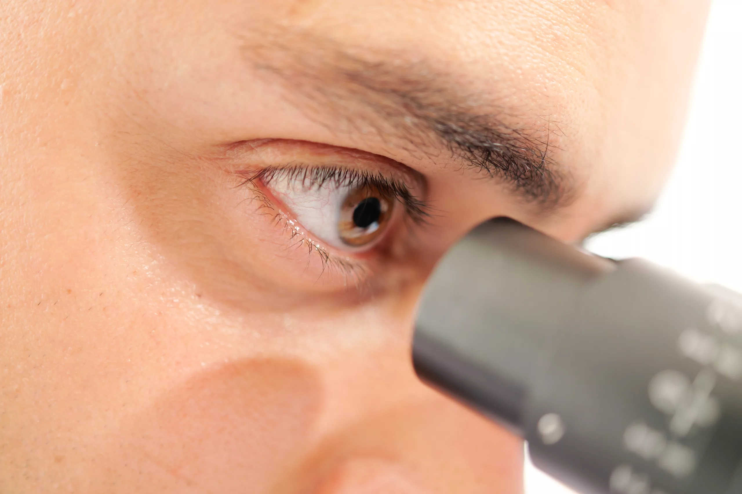
- Clinical Evaluation: A thorough physical examination may reveal palpable bony enlargement around the orbit, changes in eye position, or signs of optic nerve compression such as reduced visual acuity or color vision defects.
- Imaging Studies: X-rays, computed tomography (CT), and magnetic resonance imaging (MRI) are essential for assessing bone changes, determining the extent of orbital involvement, and identifying compression of surrounding structures, including the optic nerve and other cranial nerves.
Management
- Medical Therapy: Bisphosphonates, such as alendronate or pamidronate, are the mainstay of medical treatment for Paget’s disease. They inhibit osteoclast activity, thereby reducing bone turnover and slowing disease progression. Calcitonin may also be used in some cases to help regulate bone metabolism.
- Surgical Intervention: Surgical options may be considered in cases of severe optic nerve compression or significant proptosis that does not respond to medical therapy. Surgical decompression aims to alleviate pressure on the optic nerve and improve visual function.
- Symptomatic Treatment: Management of specific symptoms, such as dry eye syndrome due to proptosis, and supportive care to enhance quality of life.
Prognosis
- The prognosis of ocular Paget’s disease depends on various factors, including the extent of bone involvement, early diagnosis, and prompt initiation of treatment.
- With timely intervention and appropriate management, including regular monitoring of disease progression and response to therapy, complications such as optic nerve damage can be minimized, and visual function can be preserved.
Conclusion
Ocular Paget’s disease presents unique challenges due to its potential impact on vision and facial aesthetics. Early recognition, thorough evaluation, and a multidisciplinary approach involving ophthalmologists, rheumatologists, and orthopedic surgeons are essential for optimal management. Continued research into the pathophysiology and treatment of Paget’s disease aims to improve outcomes and quality of life for affected individuals, emphasizing the importance of comprehensive care tailored to individual patient needs.
World Eye Care Foundation’s eyecare.live brings you the latest information from various industry sources and experts in eye health and vision care. Please consult with your eye care provider for more general information and specific eye conditions. We do not provide any medical advice, suggestions or recommendations in any health conditions.
Commonly Asked Questions
The prognosis depends on several factors, including the severity of bone involvement, early diagnosis, and response to treatment. With appropriate management, including regular monitoring and adjustments to treatment as needed, many individuals can maintain good vision and quality of life. However, close collaboration with healthcare providers is essential to manage potential complications effectively.
Yes, ocular Paget’s disease can affect one or both eyes, although it may not necessarily affect both eyes symmetrically. Proptosis and other symptoms can vary between the eyes depending on the extent of bone involvement and compression of orbital structures.
While lifestyle changes alone cannot treat ocular Paget’s disease, maintaining a healthy lifestyle with adequate nutrition and regular exercise can support overall bone health. Avoiding smoking and excessive alcohol consumption may also be beneficial.
Yes, there is a genetic predisposition to Paget’s disease, with mutations in certain genes (like SQSTM1) increasing the likelihood of developing the condition. However, not everyone with these genetic mutations will develop Paget’s disease, as environmental factors also play a role.
Treatment aims to slow down bone turnover and manage symptoms. Bisphosphonates are commonly prescribed to inhibit osteoclast activity and reduce bone resorption. Surgical intervention may be necessary in cases of severe proptosis or optic nerve compression that does not respond to medical therapy.
In severe cases where there is significant compression of the optic nerve or other structures within the orbit, ocular Paget’s disease can lead to permanent vision loss. Early diagnosis and appropriate management are crucial in preventing irreversible damage to vision.
Diagnosis typically involves a combination of clinical evaluation and imaging studies. A physical examination may reveal palpable bony enlargement around the orbit or signs of optic nerve compression. Imaging modalities such as X-rays, CT scans, or MRI are used to visualize bone changes and assess the extent of orbital involvement.
Early signs may include mild proptosis (eye bulging), which can progress over time. Some individuals may notice changes in their vision, such as blurred vision or difficulty with eye movements. Facial asymmetry or a feeling of pressure behind the eyes can also occur.
Ocular involvement in Paget’s disease is relatively rare, occurring in a small percentage of individuals diagnosed with Paget’s disease of the bone. It tends to affect older adults more frequently and may manifest as the disease progresses.
Paget’s disease is believed to result from a combination of genetic factors and environmental triggers. Genetic mutations, particularly in genes like SQSTM1, predispose individuals to the disease. Environmental factors such as viral infections (like paramyxoviruses) may also play a role in triggering abnormal bone remodeling.


news via inbox
Subscribe here to get latest updates !


