Ocular Synechiae: Causes, Symptoms, Treatment
Embark on a journey to understand ocular synechiae, a condition characterized by adhesions between the iris and other eye structures. This article serves as your guide, providing insights into the nature of ocular synechiae, its causes, symptoms, and the importance of seeking professional help. Delve into risk factors, complications, diagnosis, treatment options, and practical measures for managing ocular synechiae and preserving eye health.
Overview of Ocular Synechiae
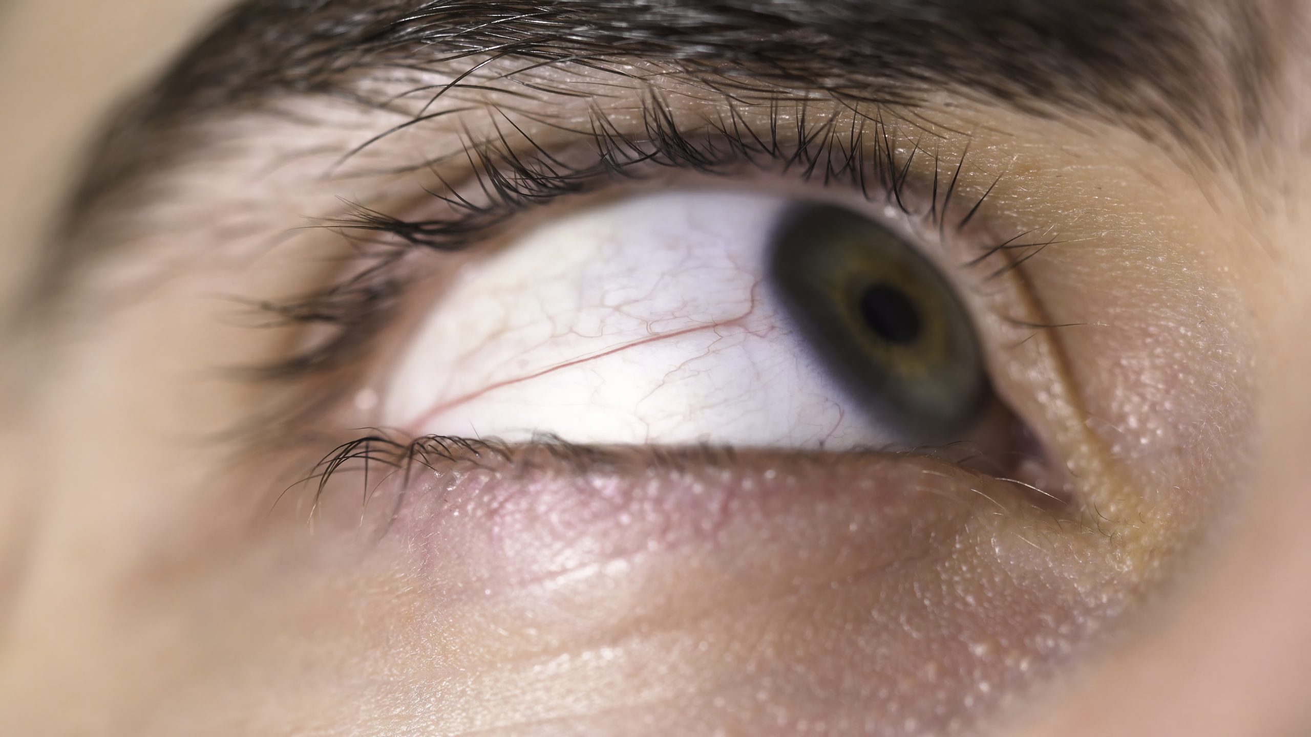
Ocular synechiae refers to adhesions or attachments between the iris (colored part of the eye) and other structures within the eye, most commonly the cornea or lens. These adhesions can limit the normal movement of the iris, leading to various complications. Ocular synechiae can be associated with conditions such as uveitis, which is inflammation of the uvea (the middle layer of the eye).
Symptoms
- Eye Pain: Individuals with ocular synechiae may experience eye pain, especially when there is inflammation or increased intraocular pressure.
- Blurred Vision: Adhesions between the iris and other eye structures can affect the normal functioning of the eye, leading to blurred or distorted vision.
- Photophobia: Sensitivity to light may be heightened, causing discomfort in well-lit environments.
- Redness: The affected eye may appear red due to underlying inflammation.
- Changes in Pupil Shape: Adhesions can alter the shape of the pupil, causing irregularities.
Causes
- Uveitis: Inflammation of the uvea, which can result from various causes, including infections, autoimmune diseases, or trauma, is a common cause of ocular synechiae.
- Posterior Synechiae: Adhesions between the iris and the lens, often seen in conditions like posterior uveitis.
- Anterior Synechiae: Adhesions between the iris and the cornea, typically associated with anterior uveitis.
- Injury or Trauma: Physical injury to the eye can lead to inflammation and the formation of synechiae.
What Happens Because of the Condition
Ocular synechiae can have several consequences, including:
- Iris Deformities: Adhesions can lead to changes in the shape and movement of the iris, affecting the regulation of pupil size.
- Glaucoma: Ocular synechiae can contribute to increased intraocular pressure, leading to glaucoma. This is particularly true if the drainage angle of the eye becomes obstructed.
- Vision Impairment: Depending on the extent and location of the synechiae, individuals may experience varying degrees of vision impairment.
Risk Factors
- Uveitis: Individuals with uveitis, especially those with chronic or recurrent uveitis, are at an increased risk of developing ocular synechiae.
- Inflammatory Conditions: Autoimmune diseases and other inflammatory conditions can contribute to the development of uveitis and subsequent synechiae.
- Eye Trauma: Injuries to the eye, such as blunt trauma or penetrating injuries, increase the risk of inflammation and adhesion formation.
- Age: While ocular synechiae can occur at any age, it may be more common in older individuals.
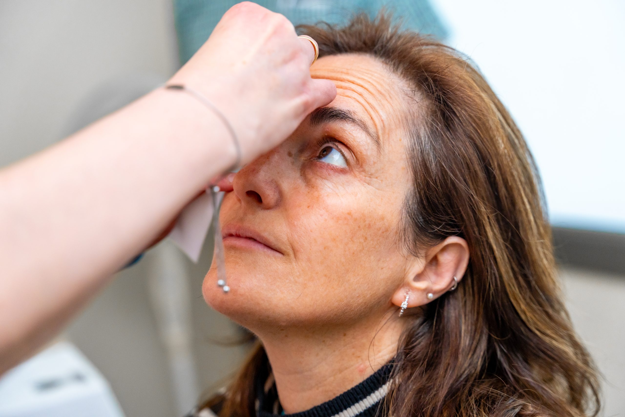
Diagnosis
- Comprehensive Eye Examination: A thorough eye examination, including visual acuity testing and slit-lamp examination, is essential for detecting ocular synechiae.
- Pupil Dilation: Dilating the pupil allows the eye care professional to assess the extent of adhesions and any associated changes in pupil shape.
- Intraocular Pressure Measurement: Elevated intraocular pressure may indicate the presence of glaucoma, which can be associated with ocular synechiae.
- Imaging Studies: In some cases, imaging studies such as ultrasound or optical coherence tomography (OCT) may be used to visualize the structures inside the eye.
Early diagnosis and appropriate management of the underlying cause, such as uveitis, are crucial for preventing complications associated with ocular synechiae. Treatment may involve anti-inflammatory medications, pupil dilation, and, in severe cases, surgical intervention to break adhesions and restore normal eye function. Regular follow-up with an eye care professional is essential to monitor the condition and adjust treatment as needed.
Treatment Options
- Anti-inflammatory Medications:
- Topical Steroids: To reduce inflammation within the eye.
- Nonsteroidal Anti-Inflammatory Drugs (NSAIDs): These medications can also help control inflammation.
- Mydriatic (Pupil Dilation) Drops:
- Atropine or Scopolamine: These medications help prevent or break adhesions by keeping the pupil dilated.
- Cycloplegic Medications:
- Cyclopentolate or Homatropine: These medications can help relax the ciliary muscle and alleviate pain associated with inflammation.
- Surgical Intervention:
- Lysis of Adhesions: In severe cases, surgical procedures may be performed to break the adhesions between the iris and other structures within the eye.
- Management of Underlying Conditions:
- Treating the underlying cause of uveitis or inflammation is crucial to preventing the formation of ocular synechiae.
Complications
- Glaucoma: Ocular synechiae can lead to impaired drainage of the aqueous humor, increasing intraocular pressure and contributing to the development of glaucoma.
- Vision Loss: Depending on the extent and location of adhesions, vision impairment can occur.
- Iris Abnormalities: The shape and movement of the iris may be altered, leading to cosmetic and functional changes.
- Recurrence: Ocular synechiae can sometimes recur, especially in cases of chronic uveitis.
Prevention
- Early Treatment of Uveitis: Prompt and effective management of uveitis reduces the risk of developing ocular synechiae.
- Regular Eye Examinations: Routine eye check-ups can help detect and address underlying conditions before they progress to complications.
- Compliance with Medications: Adherence to prescribed medications, especially anti-inflammatory and pupil-dilating drops, is crucial for preventing and managing ocular synechiae.
Medications
- Topical Steroids:
- Prednisolone: Often used to reduce inflammation in the eye.
- Nonsteroidal Anti-Inflammatory Drugs (NSAIDs):
- Ketorolac or Flurbiprofen: These medications help control intraocular inflammation.
- Mydriatic (Pupil Dilation) Drops:
- Atropine or Scopolamine: These medications prevent or break adhesions by keeping the pupil dilated.
- Cycloplegic Medications:
- Cyclopentolate or Homatropine: Used to relax the ciliary muscle and alleviate pain associated with inflammation.
When to See a Doctor
It is important to consult an eye care professional if you experience:
- Eye Pain or Discomfort: Persistent pain or discomfort, especially when accompanied by other symptoms like blurred vision or sensitivity to light.
- Changes in Vision: Any sudden changes in vision or the appearance of floaters.
- Eye Redness: Persistent redness, which may indicate underlying inflammation.
- Light Sensitivity: Increased sensitivity to light (photophobia).
- History of Uveitis or Eye Trauma: Individuals with a history of uveitis or recent eye trauma should seek prompt evaluation.
Early intervention is crucial to prevent complications and preserve vision. If you suspect ocular synechiae or experience any concerning symptoms, do not hesitate to schedule an appointment with an eye care professional for a comprehensive examination.
Demographics More Susceptible
- Individuals with Uveitis History:
- People who have a history of uveitis, which is inflammation of the uvea, are at an increased risk of developing ocular synechiae.
- Autoimmune Disease Patients:
- Individuals with autoimmune diseases, such as rheumatoid arthritis or lupus, may be more susceptible to uveitis and subsequent ocular synechiae.
- Eye Trauma Survivors:
- Those who have experienced eye injuries, including blunt trauma or penetrating injuries, are at a higher risk of developing inflammation and adhesions leading to ocular synechiae.
- Age-Related Risk:
- While ocular synechiae can occur at any age, it may be more common in older individuals who may be prone to age-related eye conditions.
- Patients with Chronic Uveitis:
- Individuals with chronic or recurrent uveitis are more likely to develop ocular synechiae, emphasizing the importance of consistent monitoring and follow-up care.
Follow-up Care for Adults and Children
- Children:
- Children diagnosed with ocular synechiae, often associated with pediatric uveitis, require diligent follow-up care.
- Pediatric ophthalmologists will monitor the extent of adhesions, assess visual acuity, and adjust treatment plans accordingly.
- Regular eye examinations are crucial to detect and manage any recurrence of uveitis or development of complications.
- Adults:
- Adults with ocular synechiae also need ongoing follow-up care with an eye care professional, typically an ophthalmologist.
- Regular examinations will include assessments of intraocular pressure, visual acuity, and the overall health of the eye.
- Adjustments to medications or additional interventions may be made based on the individual’s response to treatment and any changes in their eye health.
- Monitoring for Complications:
- Regular follow-up appointments are essential to monitor for complications such as glaucoma, changes in vision, or recurrent uveitis.
- In cases where surgical intervention has been performed, post-operative care and follow-up are crucial for assessing the success of the procedure and managing any potential issues.
- Education and Support:
- Both adults and children benefit from education about their condition and the importance of medication adherence.
- Support groups or counseling may be helpful, especially for children, to cope with the emotional aspects of living with a chronic eye condition.
Conclusion
In conclusion, ocular synechiae is a condition that requires ongoing attention and care, particularly for individuals with specific risk factors such as a history of uveitis, autoimmune diseases, or eye trauma. Diligent follow-up care is essential for both adults and children to monitor the progression of the condition, manage complications, and adjust treatment plans as needed. Timely intervention, consistent medication adherence, and regular eye examinations play pivotal roles in preserving vision and maintaining overall eye health. Through collaborative efforts between patients, their families, and eye care professionals, the impact of ocular synechiae can be minimized, leading to improved quality of life and visual outcomes.
World Eye Care Foundation’s eyecare.live brings you the latest information from various industry sources and experts in eye health and vision care. Please consult with your eye care provider for more general information and specific eye conditions. We do not provide any medical advice, suggestions or recommendations in any health conditions.
Commonly Asked Questions
Ocular synechiae can be associated with an increased risk of glaucoma, especially if the adhesions contribute to changes in intraocular pressure. Regular monitoring is essential to assess for glaucomatous changes.
Preventing ocular synechiae involves managing underlying inflammatory conditions, seeking prompt medical attention for eye symptoms, and following prescribed treatment plans to prevent recurrence.
Ocular synechiae can occur at any age, but it is more commonly associated with inflammatory eye conditions that may affect individuals of various age groups.
Ocular synechiae itself may not directly cause headaches. However, underlying inflammation or changes in intraocular pressure associated with the condition could potentially contribute to headache symptoms.
The presence of ocular synechiae may not be easily visible without specialized examination equipment. Eye care professionals use various techniques, including slit-lamp examination, to assess the extent of adhesions.
Recurrence of ocular synechiae is possible, especially if the underlying inflammatory condition persists. Regular follow-up appointments and ongoing management are essential for preventing recurrence.
Some individuals with ocular synechiae may not experience noticeable symptoms initially. Regular eye check-ups are crucial for detecting and managing the condition, even in the absence of overt symptoms.
In some cases, ocular synechiae may be associated with systemic diseases that contribute to inflammation, such as rheumatoid arthritis or lupus. A comprehensive medical evaluation may be necessary.
Ocular synechiae is relatively uncommon but can occur in individuals with inflammatory eye conditions. The prevalence may vary based on underlying risk factors.
Untreated ocular synechiae may contribute to complications such as increased intraocular pressure and optic nerve damage, potentially leading to permanent vision loss. Timely intervention is crucial.


news via inbox
Subscribe here to get latest updates !