Surgical Management of Ocular Surface Diseases
Introduction
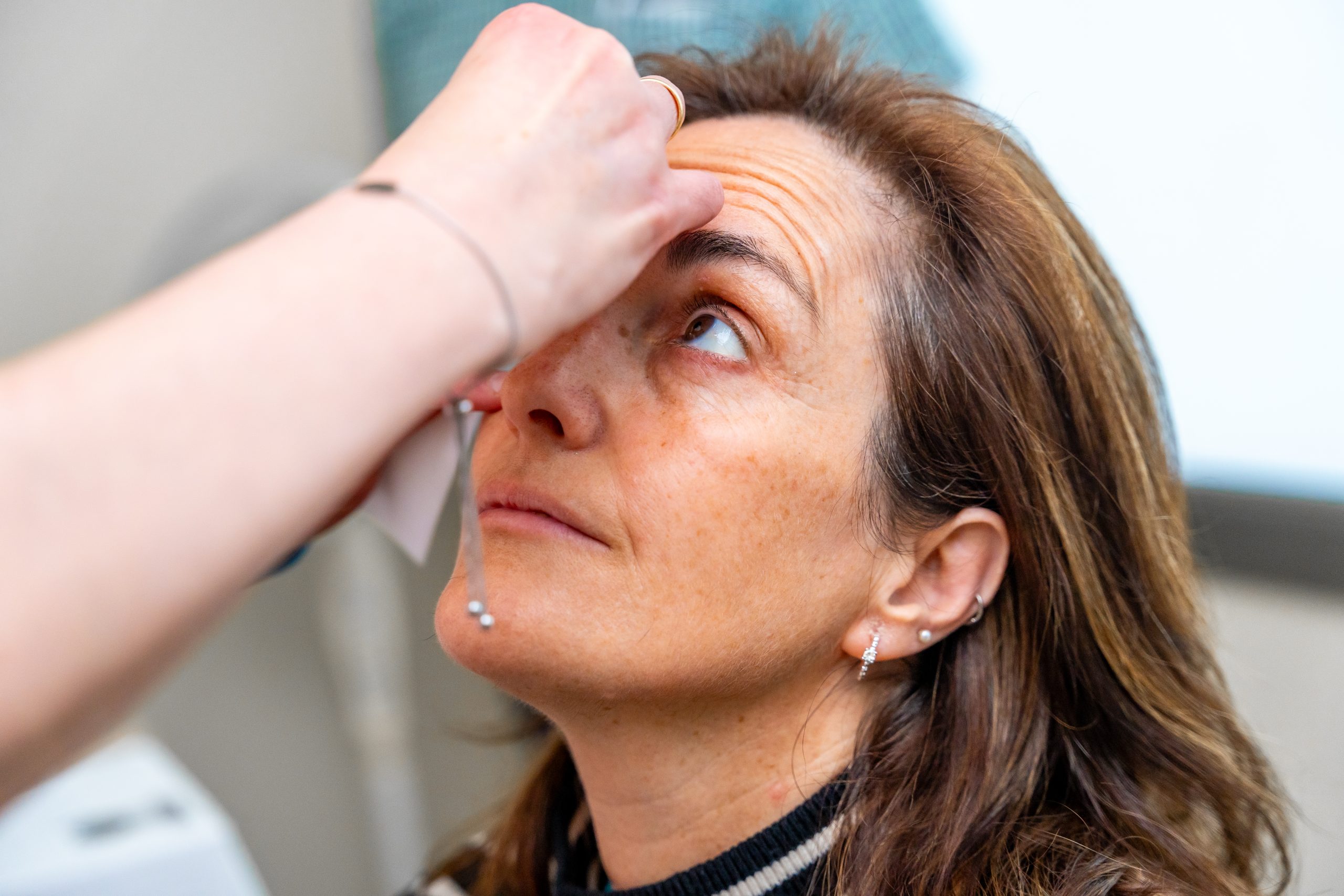
The ocular surface, comprising the cornea, conjunctiva, and associated structures, plays a crucial role in maintaining visual clarity and ocular health. Ocular surface diseases encompass a spectrum of conditions that can compromise the integrity and function of these delicate tissues, leading to discomfort, visual disturbances, and potential vision loss. While many ocular surface diseases can be managed with medical therapy, surgical interventions have emerged as valuable tools in the treatment armamentarium, offering hope for patients with refractory or advanced cases. In this article, we explore the surgical management of ocular surface diseases, highlighting innovations, techniques, and outcomes that underscore the evolving landscape of ocular surface surgery.
Common Ocular Surface Diseases
Before delving into surgical interventions, it’s essential to recognize some common ocular surface diseases:
- Dry Eye Disease: Characterized by insufficient tear production or poor tear quality, leading to ocular discomfort and visual disturbances.
- Ocular Surface Neoplasia: Including conditions such as conjunctival or corneal intraepithelial neoplasia, which may require surgical excision.
- Pterygium: A non-cancerous growth of conjunctival tissue onto the cornea, often requiring surgical removal.
- Corneal Dystrophies: Inherited disorders affecting corneal clarity and structure, sometimes necessitating corneal transplantation.
Surgical Interventions
- Limbal Stem Cell Transplantation (LSCT):
-
- Indication: LSCT is primarily indicated for patients with limbal stem cell deficiency (LSCD), a condition characterized by the loss or dysfunction of limbal stem cells responsible for regenerating the corneal epithelium.
- Techniques: LSCT techniques include autologous transplantation, where healthy limbal tissue is harvested from the patient’s unaffected eye, and allogeneic transplantation, where donor tissue is used. Additionally, ex vivo expansion of limbal epithelial cells can be performed in specialized laboratories to generate a sufficient amount of transplantable tissue.
- Procedure: The transplantation procedure involves the meticulous removal of unhealthy or scarred tissue from the affected eye’s limbal region, followed by the placement of healthy limbal tissue onto the exposed cornea. The transplanted cells then proliferate and regenerate the corneal epithelium, restoring ocular surface integrity and visual function.
- Outcomes: LSCT has shown promising outcomes in restoring vision and improving ocular comfort in patients with LSCD. Success rates vary depending on factors such as the underlying cause of LSCD, the extent of corneal involvement, and the surgical technique employed.
- Amniotic Membrane Transplantation (AMT):
-
- Indication: AMT is indicated for various ocular surface disorders characterized by epithelial defects, inflammation, or poor wound healing, such as persistent epithelial defects, corneal ulcers, chemical burns, or ocular surface reconstruction following surgical excision of neoplastic lesions.
- Techniques: AMT can be performed using dehydrated amniotic membrane grafts, cryopreserved amniotic membrane grafts, or freshly harvested amniotic membrane tissue obtained from placental donors.
- Procedure: During the procedure, the amniotic membrane graft is placed onto the affected area of the ocular surface, where it serves as a biological scaffold to promote epithelialization, reduce inflammation, and modulate wound healing. The membrane gradually dissolves over time, leaving behind a regenerated epithelial layer and improved ocular surface health.
- Outcomes: AMT has demonstrated efficacy in promoting epithelial healing, reducing inflammation, and improving ocular surface stability in various ocular surface disorders. It is well-tolerated and associated with minimal adverse effects, making it a valuable adjunctive therapy in the management of challenging cases.
- Conjunctival Reconstruction:
-
- Indication: Conjunctival reconstruction techniques are indicated for patients with extensive conjunctival scarring, neoplastic lesions, or severe dry eye disease refractory to conservative treatments.
- Techniques: Various techniques can be employed for conjunctival reconstruction, including conjunctival autografts, rotational flaps, or amniotic membrane grafting.
- Procedure: Depending on the specific indication and extent of conjunctival involvement, the surgeon may harvest healthy conjunctival tissue from a donor site on the patient’s eye or use donor tissue obtained from placental sources. The harvested tissue is then transplanted onto the affected area of the conjunctiva to restore ocular surface integrity and improve tear film stability.
- Outcomes: Conjunctival reconstruction procedures aim to alleviate symptoms of ocular discomfort, improve tear film quality, and promote ocular surface healing in patients with conjunctival scarring or dysfunction. Success rates vary depending on the underlying pathology and the surgical technique employed.
- Corneal Transplantation:
-
- Indication: Corneal transplantation, also known as keratoplasty, is indicated for patients with corneal disease or damage that cannot be adequately managed with conservative measures. Common indications include corneal scarring, keratoconus, corneal dystrophies, corneal endothelial dysfunction, or corneal perforations.
- Techniques: Different techniques of corneal transplantation are available, including penetrating keratoplasty (PKP), deep anterior lamellar keratoplasty (DALK), Descemet’s stripping automated endothelial keratoplasty (DSAEK), and Descemet’s membrane endothelial keratoplasty (DMEK), each targeting specific layers of the cornea and underlying pathology.
- Procedure: During corneal transplantation, the diseased or damaged corneal tissue is surgically removed and replaced with healthy donor corneal tissue obtained from a deceased donor. The donor tissue is carefully sutured or positioned onto the recipient’s cornea, where it integrates and restores corneal clarity and function over time.
- Outcomes: Corneal transplantation is associated with high success rates and can significantly improve visual acuity and quality of life in patients with corneal disease or injury. Advances in surgical techniques, tissue preservation, and postoperative management have contributed to improved outcomes and reduced rejection rates in corneal transplantation procedures.
Conclusion
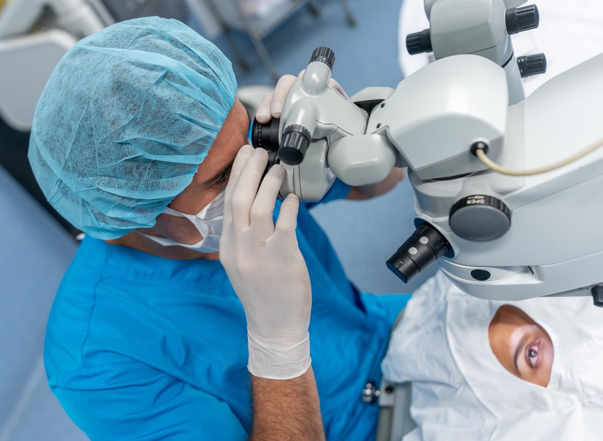
Surgical management of ocular surface diseases represents a dynamic field characterized by innovation and refinement of techniques aimed at preserving vision and improving patient outcomes. From limbal stem cell transplantation to corneal transplantation, surgical interventions offer hope for patients with challenging ocular surface conditions. As our understanding of ocular surface physiology and surgical techniques continues to evolve, the future holds promise for further advancements in the management of ocular surface diseases, ultimately enhancing the quality of life for patients worldwide.
World Eye Care Foundation’s eyecare.live brings you the latest information from various industry sources and experts in eye health and vision care. Please consult with your eye care provider for more general information and specific eye conditions. We do not provide any medical advice, suggestions or recommendations in any health conditions.
Commonly Asked Questions
Your surgeon will provide post-operative instructions tailored to your specific procedure and individual needs. These may include using prescribed medications, avoiding strenuous activities, protecting the eyes from irritants, and attending follow-up appointments for monitoring and evaluation of healing progress.
Ocular surface surgery may be performed in pediatric patients for certain conditions, such as congenital corneal abnormalities or severe ocular surface diseases. Pediatric ophthalmologists can assess eligibility and provide appropriate recommendations for surgical management.
Coverage for ocular surface surgery depends on your insurance plan, the specific procedure performed, and the medical necessity as determined by your healthcare provider. It’s essential to check with your insurance provider to understand your coverage and any potential out-of-pocket expenses.
The duration of surgical outcomes varies depending on the specific condition treated, surgical technique, and individual factors. Some procedures may provide long-lasting improvement, while others may require periodic monitoring or additional treatments over time.
During your consultation, your eye surgeon will perform a comprehensive eye examination, review your medical history, discuss treatment options, and address any questions or concerns you may have about the procedure.
Ocular surface surgery aims to improve visual function and alleviate symptoms associated with ocular surface diseases. While it may not fully restore vision in all cases, surgery can often significantly enhance visual clarity and quality of life.
In some cases, conservative treatments such as medications, lubricating eye drops, or specialty contact lenses may effectively manage ocular surface diseases. However, surgery may be necessary for advanced or refractory cases.
Recovery time varies depending on the type of surgery performed and individual healing factors. Typically, patients can expect improvement in symptoms within a few days to weeks after surgery, with full recovery taking several weeks to months.
Most ocular surface surgeries are performed under local anesthesia, so you should not feel pain during the procedure. Some discomfort or mild soreness may occur during the recovery period, which can usually be managed with medication.
Ocular surface surgery, like any surgical procedure, carries risks such as infection, inflammation, or failure of the graft to integrate properly. Your surgeon will discuss potential risks and benefits before the procedure.


news via inbox
Subscribe here to get latest updates !